Review
. 2014 Oct;66(4):948-83.
doi: 10.1124/pr.113.008201.

Bile acid signaling in metabolic disease and drug therapy

Affiliations
Review

Bile acid signaling in metabolic disease and drug therapy

Tiangang Li et al. Pharmacol Rev. 2014 Oct.

Abstract

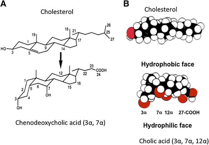
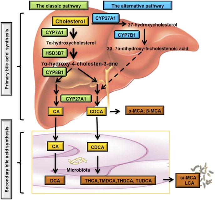
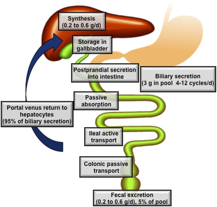
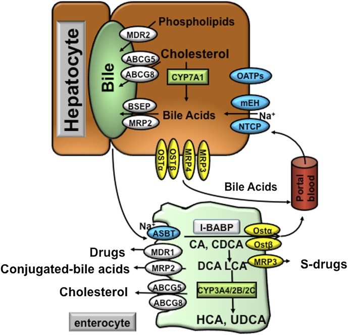
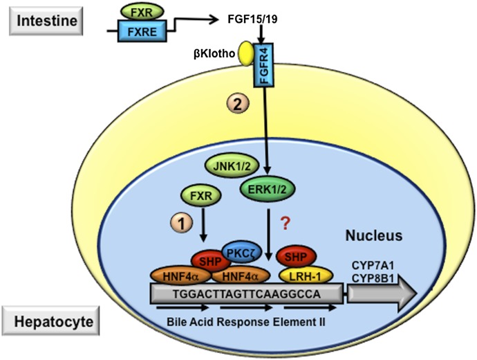
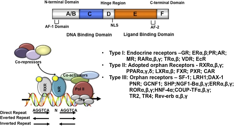
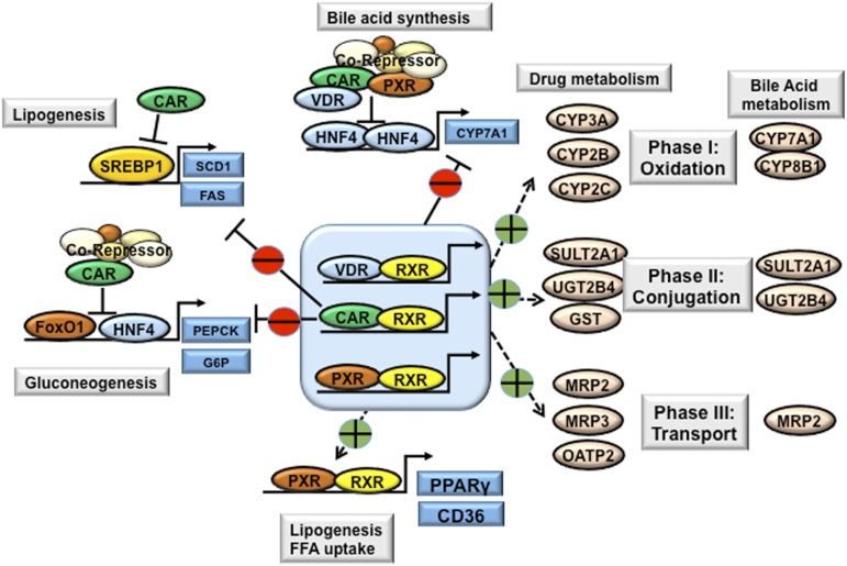
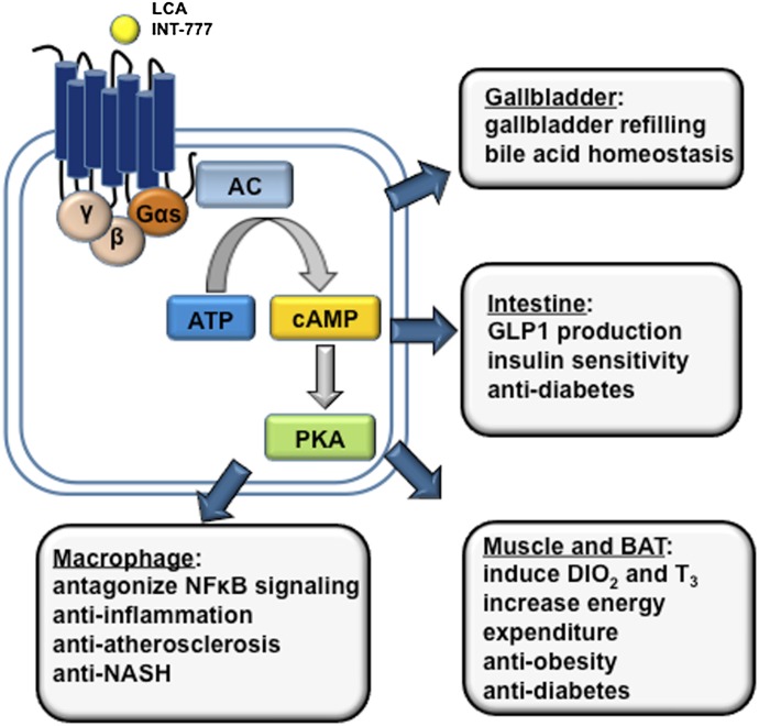
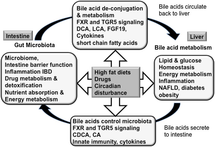
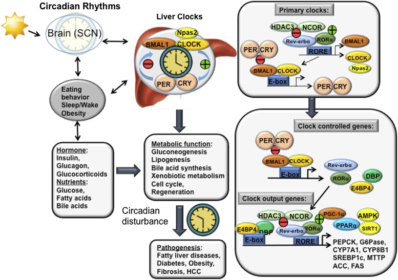
Bile acids are the end products of cholesterol catabolism. Hepatic bile acid synthesis accounts for a major fraction of daily cholesterol turnover in humans. Biliary secretion of bile acids generates bile flow and facilitates hepatobiliary secretion of lipids, lipophilic metabolites, and xenobiotics. In the intestine, bile acids are essential for the absorption, transport, and metabolism of dietary fats and lipid-soluble vitamins. Extensive research in the last 2 decades has unveiled new functions of bile acids as signaling molecules and metabolic integrators. The bile acid-activated nuclear receptors farnesoid X receptor, pregnane X receptor, constitutive androstane receptor, vitamin D receptor, and G protein-coupled bile acid receptor play critical roles in the regulation of lipid, glucose, and energy metabolism, inflammation, and drug metabolism and detoxification. Bile acid synthesis exhibits a strong diurnal rhythm, which is entrained by fasting and refeeding as well as nutrient status and plays an important role for maintaining metabolic homeostasis. Recent research revealed an interaction of liver bile acids and gut microbiota in the regulation of liver metabolism. Circadian disturbance and altered gut microbiota contribute to the pathogenesis of liver diseases, inflammatory bowel diseases, nonalcoholic fatty liver disease, diabetes, and obesity. Bile acids and their derivatives are potential therapeutic agents for treating metabolic diseases of the liver.


Figures

Fig. 1.
Fig. 1.
Bile acid structure. (A) Conversion of cholesterol to bile acid alters the stereo-configuration of the steroid ring structure. Saturation of the C5-C6 double bond changed the hydrogen group in C5 from α to β and a cis-configuration along the plane of the fused A and B ring, and caused a kink along the steroid plane in CDCA. (B) Space-filling models of cholesterol and CA. All three hydroxyl groups and the carboxyl group are faced to one side of the carbon skeleton to form a hydrophilic face, which is opposite to the hydrophobic face of the carbon skeleton.
Fig. 2.
Fig. 2.
Bile acid biosynthetic pathways. Two major bile acid biosynthetic pathways are shown. In the classic pathway, cholesterol is converted to 7α-hydroxycholesterol by the rate-limiting enzyme CYP7A1, which is located in the ER. The 3β-hydroxysteroid dehydrogenase (3βHSD, HSD3B7) converts 7α-hydroxycholesterol to 7α-hydroxy-4-cholesten-3-one (C4), which is converted to 7α,12α-dihydroxy-4-cholesten-3-one by a sterol 12α-hydroxylase (CYP8B1) leading to synthesis of CA. Without 12α-hydroxylation by CYP8B1, C4 is eventually converted to CDCA. The mitochondrial sterol 27-hydroxylase (CYP27A1) catalyzes the steroid side chain oxidation in both CA and CDCA synthesis. In the alternative pathway, cholesterol is first converted to 27-hydroxycholesterol by CYP27A1. Oxysterol 7α- hydroxylase (CYP7B1) catalyzes hydroxylation of 27-hydroxycholesterol to 3β,7α-dihydroxy-5-cholestenoic acid, which eventually is converted to CDCA. Oxysterol 7α-hydroxylase (CYP7B1) is nonspecific and can also catalyze hydroxylation of 25-hydroxycholesterol to 5-cholesten-3β,7α,25-triol. In the large intestine, bacterial 7α-dehydroxylase removes a hydroxyl group from C-7 and converts CA to DCA and CDCA to LCA. In mouse liver, most of CDCA is converted to α- and β-MCA. In the intestine, bacterial 7α-dehydroxylase activity convers CA and CDCA to DCA and LCA, respectively. CYP3A1 and epimerase also convert CDCA to the secondary bile acids, including THCA, TMDCA, ω-MCA, THDCA, and TUDCA. LCA and ω-MCA are excreted into feces. THCA, taurohyocholic acid; THDCA, taurohyodeoxycholic acid; TMDCA, tauromurideoxycholic acid.
Fig. 3.
Fig. 3.
Enterohepatic circulation of bile acids. The human bile acid pool consists of approximately 3 g of bile acids. Food intake stimulates the gallbladder to release bile acids into the small intestine. An average man produces approximately 0.5 g bile acid per day by synthesis in the liver, and secretes approximately 0.5 g/day. Conjugated bile acids are efficiently reabsorbed in the ileum by active transport, whereas a small amount of unconjugated bile acids is reabsorbed by passive diffusion in the small and large intestines. The first-pass extraction of bile acids from the portal blood by the liver is very efficient. Small amounts of bile acids that spilled over into the systemic circulation are recovered in kidney. The bile acids in the pool are recycled 4–12 times a day.
Fig. 4.
Fig. 4.
Bile acid transporters in the hepatocytes and enterocytes. At the basolateral membrane of the hepatocytes, the NTCP and mEH may be responsible for Na+-dependent uptake of conjugated bile acids, whereas OATPs show substrate specificity for unconjugated bile acids. At the canalicular membrane of the hepatocytes, the BSEP plays a major role in biliary secretion of bile acids, whereas the MRP2 mediates secretion of organic substrates including bile acids, bilirubin, and glutathione. ABCG5 and ABCG8 heterodimers transport cholesterol into the bile, whereas MDR2 is responsible for biliary secretion of phospholipids. At the basolateral membrane of the hepatocytes, organic solute transporters OSTα and OSTβ heterodimers, MRP3, and MRP4 mediate secretion of bile acids into the circulation. With cholestasis, both basolateral bile acid efflux and renal bile acid excretion are increased. After bile acids are released from the gallbladder into the intestine, ileal bile acid uptake is mediated by the ASBT. Intracellular bile acids are bound to the intestinal bile acid binding protein (IBABP). At the basolateral membrane, bile acid efflux is mediated by the OSTα and OSTβ heterodimers. At the apical membrane of the enterocytes, ABCG5 and ABCG8 heterodimers transport cholesterol back into the intestinal lumen, a process that limits intestine cholesterol absorption. CYP3A4, CYP2B, and CYP2C are involved in the metabolism and detoxification of LCA in the intestine. In the apical membrane of intestine, MDR1 effluxes drugs and MRP2 effluxes conjugated bile acids. In the sinusoidal membrane, MRP3 effluxes sulfur-conjugated drugs for renal excretion.
Fig. 5.
Fig. 5.
Mechanisms of bile acid feedback inhibition of bile acid synthesis. Bile acid–activated signaling inhibits CYP7A1 and CYP8B1 and therefore reduces hepatic bile acid synthesis. The bile acid response element (BARE) located in the CYP7A1 gene promoter contains AGGTCA-like direct repeats. HNF4α and LRH1 bind to the BARE and stimulate CYP7A1 gene transcription. In hepatocytes, bile acids activate FXR, which induces the repressor SHP. SHP interacts with and represses the transactivating action of HNF4α and LRH, a process that involves the recruitment of corepressor complex and chromatin remodeling enzymes (indicated as pathway 1). In the intestine, bile acid–activated FXR induces FGF15 (FGF19 in humans), which binds and activates FGFR4 on the hepatocytes. FGFR4 activates intracellular signaling pathways, such as ERK, protein kinase Cζ (PKCζ), and c-Jun N-terminal kinase (JNK), which leads to the repression of CYP7A1 gene transcription (indicated in pathway 2).
Fig. 6.
Fig. 6.
Nuclear receptors. The domain structure of nuclear receptors is shown on the top. The putative nuclear receptor response element binding sequences, arranged in direct repeat, everted repeat, and inverted repeat, are shown. Ligand-activated receptors recruit coactivators to replace corepressors, which results in transactivation of target gene expression. Nuclear receptors are classified into three types: type I endocrine receptors, type II adapted orphan receptors, and type III orphan receptors. Refer to Chawla et al. (2001) for details on the nuclear receptor superfamily and nomenclature. AF-1, activation function-1. AF-2, activation function-2; NLS, nuclear localization sequence.
Fig. 7.
Fig. 7.
Xenobiotic nuclear receptors in bile acid, drug, lipid, and glucose metabolism. The xenobiotic nuclear receptors PXR and CAR are highly expressed in the liver and intestine. VDR is highly expressed in the intestine, but is expressed at low levels in the liver. Activation of xenobiotic nuclear receptors by drugs, bile acids, and xenobiotics induces a network of genes involved in phase I, phase II, and phase III drug and bile acid metabolism and detoxification. CAR, PXR, and VDR inhibit CYP7A1 and thus bile acid synthesis via interaction with HNF4α and inhibition of HNF4α transactivation of CYP7A1. Similarly, CAR inhibits PEPCK and G6Pase involved in gluconeogenesis and inhibits SREBP-1c in lipogenesis. Activation of CAR decreases plasma glucose levels and improves hepatic steatosis in obesity and diabetes. By contrast, activation of PXR induces hepatic expression of PPARγ and CD36, leading to hepatic steatosis. FFA, free fatty acid; FoxO1, forkhead box protein O1; SCD1, stearoyl-CoA desaturase-1.
Fig. 8.
Fig. 8.
Bile acid–activated TGR5 signaling in metabolism and inflammation. TGR5 is activated by secondary LCA and synthetic agonists (e.g., INT-777). TGR5 is a Gαs GPCR that induces cAMP/PKA signaling. TGR5 is expressed in brown adipocytes, macrophages/monocytes and hepatic Kupffer cells, gallbladder epithelium, and intestine, with high levels found in the colon. TGR5 is not expressed in hepatocytes. In brown adipose tissue, TGR5 activation stimulates energy expenditure; in the intestine, TGR5 activation stimulates GLP-1 production from L cells. These metabolic effects underlie the antiobesity and antidiabetic properties of TGR5 agonists. TGR5 activation shows anti-inflammatory effects, and TGR5 activation may protect against colitis, Crohn’s disease, and atherosclerosis. TGR5 in the gallbladder epithelium regulates gallbladder refilling. AC, adenylate cyclase; DIO2, type 2 deiodinase; PKA, protein kinase A; T3, 3,5,3′-triiodothyronine.
Fig. 9.
Fig. 9.
Bile acid and gut microbiota. In the intestine, bacteria overgrowth damages intestine barrier function and causes IBD, diarrhea, and impaired drug metabolism, detoxification, and absorption. Bile acids control gut bacteria overgrowth and protect against inflammation. Gut microbiota also play a role in biotransformation of bile acids and affected bile acid composition and metabolism via FXR and TGR5 signaling in the liver. In the liver, high levels of bile acids cause liver injury. Bile acids also have anti-inflammatory functions by activating FXR and TGR5 signaling in hepatocytes to protect against metabolic diseases such as NAFLD, diabetes, and obesity. High-fat diets and drugs alter bile acid biotransformation and gut microbiota, and contribute to pathogenesis of intestinal inflammatory disease and liver-related metabolic diseases.
Fig. 10.
Fig. 10.
Circadian rhythms in liver metabolism. The central clock in the SCN synchronizes with the peripheral clock to regulate liver metabolisms. Eating behavior, sleep/wake cycle, and obesity affect central clock and liver clock functions and their synchronization. Hormones such as insulin, glucagon, and glucocorticoids, and nutrients including glucose, fatty acids, and bile acids affect circadian rhythms and liver metabolism. Bmal1 and Clock are primary clock products that bind to the E-box sequences in the Per and Cry gene promoters. Per and Cry complexes inhibit the Bmal/Clock complex in a negative loop to inhibit Per and Cry transcription. Bmal1 and Clock (also Npas2) are regulated by a negative regulator Rev-erbα, and positive regulator RORα, which bind to the same ROR response element (RORE) in the promoters. Rev-erbα recruits HDAC3 and NcoR to inhibit gene transcription and ultimately the circadian rhythms of many CCGs, such as PEPCK and G6Pase in gluconeogenesis, CYP7A1 and CYP8B1 in bile acid synthesis, and SREBP-1c and MTTP in lipogenesis in the liver. Alteration in synchronization of the central clock and liver clock contributes to the pathogenesis of fatty liver diseases, diabetes, and obesity, as well as fibrosis and hepatocellular carcinoma. HCC, hepatocellular carcinoma; MTTP, microsomal triglyceride transfer protein; NCOR, nuclear receptor corepressor; SCN, suprachiasmatic nucleus.

References

    1. Abifadel M, Varret M, Rabès JP, Allard D, Ouguerram K, Devillers M, Cruaud C, Benjannet S, Wickham L, Erlich D, et al. (2003) Mutations in PCSK9 cause autosomal dominant hypercholesterolemia. Nat Genet 34:154–156 - PubMed
    1. Adada M, Canals D, Hannun YA, Obeid LM. (2013) Sphingosine-1-phosphate receptor 2. FEBS J 280:6354–6366 - PMC - PubMed
    1. Adorini L, Pruzanski M, Shapiro D. (2012) Farnesoid X receptor targeting to treat nonalcoholic steatohepatitis. Drug Discov Today 17:988–997 - PubMed
    1. Aldridge MA, Ito MK. (2001) Colesevelam hydrochloride: a novel bile acid-binding resin. Ann Pharmacother 35:898–907 - PubMed
    1. Alemi F, Kwon E, Poole DP, Lieu T, Lyo V, Cattaruzza F, Cevikbas F, Steinhoff M, Nassini R, Materazzi S, et al. (2013a) The TGR5 receptor mediates bile acid-induced itch and analgesia. J Clin Invest 123:1513–1530 - PMC - PubMed

MeSH terms