. 2014 Nov;13(11):3164-76.
doi: 10.1074/mcp.M114.041103. Epub 2014 Jul 29.

Copper-catalyzed azide-alkyne cycloaddition (click chemistry)-based detection of global pathogen-host AMPylation on self-assembled human protein microarrays

Affiliations

Copper-catalyzed azide-alkyne cycloaddition (click chemistry)-based detection of global pathogen-host AMPylation on self-assembled human protein microarrays

Xiaobo Yu et al. Mol Cell Proteomics. 2014 Nov.

Abstract

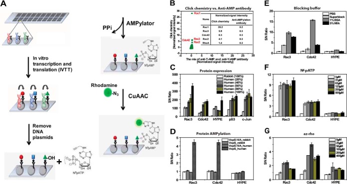
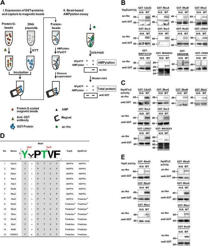
AMPylation (adenylylation) is a recently discovered mechanism employed by infectious bacteria to regulate host cell signaling. However, despite significant effort, only a few host targets have been identified, limiting our understanding of how these pathogens exploit this mechanism to control host cells. Accordingly, we developed a novel nonradioactive AMPylation screening platform using high-density cell-free protein microarrays displaying human proteins produced by human translational machinery. We screened 10,000 unique human proteins with Vibrio parahaemolyticus VopS and Histophilus somni IbpAFic2, and identified many new AMPylation substrates. Two of these, Rac2, and Rac3, were confirmed in vivo as bona fide substrates during infection with Vibrio parahaemolyticus. We also mapped the site of AMPylation of a non-GTPase substrate, LyGDI, to threonine 51, in a region regulated by Src kinase, and demonstrated that AMPylation prevented its phosphorylation by Src. Our results greatly expanded the repertoire of potential host substrates for bacterial AMPylators, determined their recognition motif, and revealed the first pathogen-host interaction AMPylation network. This approach can be extended to identify novel substrates of AMPylators with different domains or in different species and readily adapted for other post-translational modifications.


Figures

Fig. 1.
Fig. 1.
Development of AMPylation screening platform on NAPPA arrays. A, The principle of AMPylation assay on NAPPA arrays. B, Comparison of AMPylation assay on NAPPA arrays using click chemistry and anti-AMPylation antibody. The experiment was performed by using VopS enzyme and NAPPA GST2 arrays comprising 1823 human proteins. 1:50 dilution of anti-T-AMP antibody and anti-Y-AMP antibody were mixed and used as the primary antibody, and 1:100 dilution of Alex647 chick anti-rabbit IgG antibody was used as the secondary detection antibody. X-axis and Y-axis represent the normalized signal intensity produced by taking the signal intensity for each feature divided by the median background-adjusted value for all the proteins on the array as described in the methods. C–G, Optimization of the assay parameters for AMPylation assay on NAPPA arrays, including the cell-free expression system C, D, blocking buffer E, N6pATP F, and az-rho G, respectively. The signal to noise ratio (S/N) ratio was calculated using MSP-1, which expresses a 25 kDa protein with the HA tag and is not captured to the anti-GST coated array surface, as a negative control. CuAAC, copper-catalyzed azide-alkyne cycloaddition.
Fig. 2.
Fig. 2.
Global identification of substrates for VopS and IbpAFic2 using high-density NAPPA arrays. A, Representative images show the quality control of fabricated high-density NAPPA arrays. Expression clones (n = 1823) encoding the target proteins fused to a C-terminal GST tag were printed along with a polyclonal anti-GST antibody in single spot on the array surface. DNA capture was confirmed by PicoGreen staining (Fig. 2A, left). The plasmid DNA was removed using DNase after expression (Fig. 2A, middle), and the protein displayed in situ was assessed by a monoclonal anti-GST antibody (Fig. 2A, right) (GST color code: red>orange>yellow>green>blue). B, The PicoGreen signal was quantified on the slide and displayed as a box (25th–75th percentiles) and whisker plot both before (Green) and after (Black) DNase treatment. MFI is the abbreviation of mean of fluorescence intensity. C, The correlation coefficient of GST signal between two NAPPA protein arrays is 0.93. D, Representative fluorescent images show human targets with strong fluorescent signals. E, F, Histograms show the distribution of the molecular weights of identified substrates for VopS and IbpAFic2 as well as the entire human 10k protein collection respectively. X-axis is the molecular weight and y axis is the number of substrate proteins. IVTT, in vitro transcription and translation.
Fig. 3.
Fig. 3.
Validation of identified human substrates from NAPPA screens. A, Schematic illustration of bead-based AMPylation assays. B, C, Validation of identified human substrates for VopS and IbpAFic2, respectively. VopS H348A and IbpAFic2 H359A, both inactive enzymes, were used as their corresponding negative controls. CD48 is an example of a substrate candidate that did not validate in the bead-based assay. D, Prediction of new substrates for VopS and IbpAFic2 by using motif analysis. In Rac1, RhoA, and Cdc42, the conserved Y and T indicated were reported as the AMPylation substrate of IbpA and VopS respectively (2, 49). Additional candidate substrates with the motif were searched in the UniProtKB/Swiss-Prot human database (http://prosite.expasy.org/scanprosite/). E, Validation of predicted substrates using bead-based AMPylation assays. Each experiment was repeated three times on independent days. az-rho, azide-rhodamine; WB, Western blot.
Fig. 4.
Fig. 4.
VopS AMPylates all Rac proteins during infection. A, Schematic of the V. para infection assay with identified substrates. Cells are transfected with substrate candidate and infected with V. para. Candidate substrates are immunoprecipitated from the lysate and blotted for AMPylation. WT is V. para CAB strain which was generated from POR1 (RIMD 2210633 ΔtdhAS). The CabS strain contains a deletion for the transcriptional regulator VtrA to prevent expression of the second type three secretion system, as well as deletions of the effectors VopQ, VopR, and VPA0450 to leave VopS as the only known T3SS effector expressed and secreted. Δ and + are the CabSΔS strain with the deletion of VopS and complemented with a pBAD-VopS expression plasmid, respectively. B, Transfected 3xHA tagged Rac1, Rac2, and Rac3 were immunoprecipitated from infected cells and immunoblotted for AMPylation.
Fig. 5.
Fig. 5.
AMPylation of LyGDI on residue threonine 51 inhibits phosphorylation by Src. A, Validation of LyGDI AMPylation using anti-threonine and anti-tyrosine AMPylation antibodies. His-RhoGDI and His-LyGDI proteins were expressed in E. coli with GST-NΔ30VopS or empty vector and blotted for total protein (anti-His), threonine AMPylation (anti-T-AMP), tyrosine AMPylation (anti-Y-AMP), and VopS expression (anti-VopS, total lysate). Lane 1 (*) is tyrosine AMPylated Cdc42. B, AMPylated or unAMPylated LyGDI incubated with Src kinase, 100 μm ATP and 5 μCi [γ-32P] ATP for 45 min. CB, coomassie staining. C, Relative phosphorylation of AMPylated versus unAMPylated LyGDI over three experiments. p = 0.034.
Fig. 6.
Fig. 6.
Bacteria-human interactions through AMPylation. A, Bacteria-human interaction AMPylation network. B, C, Distribution of PANTHER protein classes and sub-cellular location of VopS and IbpAFic2 substrates.
Fig. 7.
Fig. 7.
Biological processes of VopS and IbpAFic2 involved in the human host by AMPylation of their substrates. In both panels, GO biological processes are represented in a tree format that shows the hierarchy of GO terms. The adjusted p value from enrichment tests is represented by the color from yellow (5 × 10⁻²) to orange (5 × 10⁻⁷) (details in Methods, full-scale images in Supplemental Fig. S10 and S11 with details in supplemental Tables S5, S6) of identified substrates for each biological process.

References

    1. Muller M. P., Peters H., Blumer J., Blankenfeldt W., Goody R. S., Itzen A. (2010) The Legionella effector protein DrrA AMPylates the membrane traffic regulator Rab1b. Science 329, 946–949 - PubMed
    1. Yarbrough M. L., Li Y., Kinch L. N., Grishin N. V., Ball H. L., Orth K. (2009) AMPylation of Rho GTPases by Vibrio VopS disrupts effector binding and downstream signaling. Science 323, 269–272 - PubMed
    1. Worby C. A., Mattoo S., Kruger R. P., Corbeil L. B., Koller A., Mendez J. C., Zekarias B., Lazar C., Dixon J. E. (2009) The fic domain: regulation of cell signaling by adenylylation. Mol. Cell 34, 93–103 - PMC - PubMed
    1. Neunuebel M. R., Chen Y., Gaspar A. H., Backlund P. S., Jr., Yergey A., Machner M. P. (2011) De-AMPylation of the small GTPase Rab1 by the pathogen Legionella pneumophila. Science 333, 453–456 - PMC - PubMed
    1. Tan Y., Luo Z. Q. (2011) Legionella pneumophila SidD is a deAMPylase that modifies Rab1. Nature 475, 506–509 - PMC - PubMed

Publication types

MeSH terms

LinkOut - more resources