. 2014 Dec;15(6):975-92.
doi: 10.1007/s10162-014-0480-x. Epub 2014 Jul 30.

How to bury the dead: elimination of apoptotic hair cells from the hearing organ of the mouse

Affiliations

How to bury the dead: elimination of apoptotic hair cells from the hearing organ of the mouse

Tommi Anttonen et al. J Assoc Res Otolaryngol. 2014 Dec.

Abstract

Hair cell death is a major cause of hearing impairment. Preservation of surface barrier upon hair cell loss is critical to prevent leakage of potassium-rich endolymph into the organ of Corti and to prevent expansion of cellular damage. Understanding of wound healing in this cytoarchitecturally complex organ requires ultrastructural 3D visualization. Powered by the serial block-face scanning electron microscopy, we penetrate into the cell biological mechanisms in the acute response of outer hair cells and glial-like Deiters' cells to ototoxic trauma in vivo. We show that Deiters' cells function as phagocytes. Upon trauma, their phalangeal processes swell and the resulting close cellular contacts allow engulfment of apoptotic cell debris. Apical domains of dying hair cells are eliminated from the inner ear sensory epithelia, an event thought to depend on supporting cells' actomyosin contractile activity. We show that in the case of apoptotic outer hair cells of the organ of Corti, elimination of their apices is preceded by strong cell body shrinkage, emphasizing the role of the dying cell itself in the cleavage. Our data reveal that the resealing of epithelial surface by junctional extensions of Deiters' cells is dynamically reinforced by newly polymerized F-actin belts. By analyzing Cdc42-inactivated Deiters' cells with defects in actin dynamics and surface closure, we show that compromised barrier integrity shifts hair cell death from apoptosis to necrosis and leads to expanded hair cell and nerve fiber damage. Our results have implications concerning therapeutic protective and regenerative interventions, because both interventions should maintain barrier integrity.


Figures
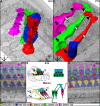
FIG. 1
FIG. 1
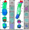
Cytoarchitectural development of the organ of Corti. AD SBEM imaged volumes with 3D-modeled OHCs and DCs of the organ of Corti at E18 (A) and P10 (C). Bending of DC phalangeal processes (open arrows) early postnatally creates distinct apical (arrowheads) and basal domains (arrows), which contact different OHCs. The basal domain of DCs differentiates into a cup-like structure. Surface views (B, D) of the imaged volumes reveal the spatial relationship between OHCs and DCs. The surface of a DC that basally contacts an OHC and the surface of this OHC are marked by identically colored spheres. E Schematic cross-sections through the organ of Corti at birth and adulthood. Schematic representations of developmental changes in DC morphology and the contact areas between these cells and OHCs. Abbreviations: T tunnel of Corti, N space of Nuel, IHC inner hair cell, IP inner pillar cell, He Hensen’s cell. Scale bars, 10 μm.
FIG. 2
FIG. 2
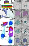
Morphological maturation of outer hair cells. A, B Side, topdown and downtop views of 3D modeled E18 (A) and P22 (B) OHCs from SBEM datasets. In the apical domain, note the increase in the length of stereocilia, formation of the cuticular plate, and disappearance of the primary cilium, the kinocilium, by adulthood. Note also the relocalization and shape changes of mitochondria along maturation. In the basal domain, the amount of mitochondria beneath the nucleus decreases by adulthood. CG, HL Single images of the respective hair cells at the plane parallel to the apical surface. Section levels are indicated by arrows in (A, B). Note the stereociliary bundle maturation (C, H), cuticular plate formation (D, I), change of the OHC circumferencial shape from pentagonal shield like to round or rounded triangle (E, F, J, K), central-to-lateral relocation of mitochondria and tubular-to-round change in mitochondrial shape (D, E, F, J, K), and appearance of open spaces between OHC lateral plasma membranes (E, F, J, K), except at the level of the nucleus (G, L). Scale bar, 5 μm.
FIG. 3
FIG. 3
Outer hair cell apoptosis following ototoxic trauma. A Schematic surface views of nontraumatized and traumatized organ of Corti, and the nomenclature used for the lesion sites. B, B’ Hematoxylin-stained paraffin section through a traumatized cochlea (B). Adjacent section shows that NKCC1 is not expressed in the organ of Corti but is found in the laterally located supporting cells, the Claudius cells, and in the stria vascularis (B’). CG, HL Single images at different heights of OHCs undergoing early and advanced apoptotic degeneration at the acute lesion site. Corresponding heights are marked by arrows in (M). Gradual shrinkage of the OHC body, mitochondrial delocalization from beneath the plasma membrane (arrowhead in F), and their degradation (K) are seen. Also, DC swelling and the resulting filling of the interstitial spaces is evident. M Side, topdown and downtop views of a 3D-modeled OHC (P22 + 36 h post-trauma) undergoing advanced apoptotic degeneration show prominent cell body shrinkage, but a largely intact apical domain (compared with Fig. 2B). Minor fusion of stereocilia can be seen (open arrows in H, M). Abbreviations: SV stria vascularis, OC organ of Corti, Cl Claudius cells, T tunnel of Corti. Scale bar, 5 μm.
FIG. 4
FIG. 4
Elimination of outer hair cells within the organ of Corti. A The nontraumatized epithelium shows distinct interstitial spaces between OHC’s lateral plasma membranes. B, C Two images from the same orientation showing swelling of the DC phalanges at the acute lesion site and filling of interstitial spaces. No indications of protrusive activity by DCs can be seen at the pericuticular level (thin arrows in AC). Note that in (B), the OHC on the right side appears to be in two portions, but in fact its intact middle portion bends out from the image plane, showing the importance of 3D analysis for reliable documentation. Apoptotic body-like debris and a degrading nucleus (arrows) are seen inside the swollen DC phalanges. D A higher magnification view of these engulfed apoptotic cell particles (arrows). In addition, apoptotic bodies waiting to be internalized are seen between swollen DC phalanges (arrowhead). E At the late lesion site, lumenal spaces re-open along with partial recovery of the volume of DC phalanges. FH Lumenal re-opening at the late lesion site is also evident in sections viewed at the plane parallel to the epithelial surface. The OHC rows are numbered. Necrotic-like cell debris (asterisks) persists in lumens. Note the OHC (nucleus marked by open arrow) of the first row that is hanging free inside the lumen. The inset shows this cell in another orientation (G). Scale bar, 5 μm.
FIG. 5
FIG. 5
Rare outer hair cells persist as bundleless cells after lesion. A Overview of the late lesion site where a bundleless OHC (blue) was found. Note also necrotic-like debris in a Deiters’ cell cup (asterisk) in the proximity to the bundleless OHC. B A single section shows that this cell is innervated by nerve fibers (arrow), demonstrating that it is not a transdifferentiated supporting cell. C A 3D reconstruction of bundleless OHC. Note the collapsed appearance of the cell body and the mitochondria with no overt morphological changes. D A single image section showing necrotic-like cell debris in the Deiters’ cell cup. This debris might originate from the bundleless OHC degenerating through secondary necrosis. Scale bar, 5 μm.
FIG. 6
FIG. 6
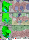
Closure of the Deiters’ cell cup and loss of synaptic terminals following ototoxic lesion. 3D models and corresponding single sections of the DC cup and associated synaptic terminals in an undamaged specimen (A, B) and at the acute (C, D) and recovery (E, F) lesion sites. While the DC cup normally fully enfolds the OHC base and clusters nerve terminals, the cup closes at the acute lesion site and loses synaptic contacts. This closure starts after detachment of the dying OHC. Note the dissociation of the cell membrane from the degrading actin plaque (asterisk in D). Note also progressive straightening of the phalangeal process (compare A and E). When the cup closure is complete at the recovery site, nerve terminals are lost and the cup’s actin plaque has disappeared (F). Open arrows mark the electron-dense F-actin plaque. Scale bar, 5 μm.
FIG. 7
FIG. 7
Cytoskeletal changes in Deiters’ cells and alterations in innervation density to outer hair cells upon ototoxic trauma, as revealed by confocal microscopy. Confocal Z-stack projections of DC cups from a nontraumatized specimen (A, A”) and from acute (B, B”) and late (C, C”) lesion sites show merged and single z-projections of phalloidin, synapsin-1 and DAPI labeling in the basal domain. Merged images (AC) show the loss of synapsin-1-immunostained nerve terminals, evident already at the acute lesion site. Note the prominent actin plaque (arrowheads) of DC cups in the control specimen and the gradual degradation of the plaque upon trauma (A’–C’). A part of DAPI-labeled OHC nuclei are shrunken and fragmented (open arrows), indicating a late stage of apoptosis (A”–C”). DF Confocal Z-stack projections of phalloidin-labeled reticular lamina show the apical domain. The dynamic remodeling and reinforcement of new surface extensions by F-actin at the site of lost OHC can be seen by comparing the intensity of phalloidin labeling at acute and late lesion sites. Insets show these events on single surface extension sites. Note also the progressive flattening of the sensory epithelium and collapse of supporting cells (arrows in B’, C’). Abbreviation: He Hensen’s cell, OHC outer hair cell, DC Deiters’ cell. Scale bar, 10 μm.
FIG. 8
FIG. 8
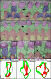
Epithelial surface closure is reinforced by F-actin belts. A, B Surface rendering from the acute lesion site shows variation in the geometry of DC junctions (arrows) that close the sites of lost OHCs (A). This variation is minimal at the late lesion site, showing that dynamic junctional remodeling takes place after the initial surface closure (B). CE’ Single block-face images and 3D models of DCs of the second row from an undamaged specimen (C, C’) and from the acute (D, D’) and late (E, E’) lesion sites. In the undamaged specimen, note that apical junctions bend at some sites and are not perpendicular to the epithelial surface. The frames of apical extensions formed upon hair cell loss are depicted (C, C’). At the acute lesion site, junctional extensions reach the sites of lost OHCs but F-actin recruitment to these extensions is minimal. The original F-actin belt is distinct (D, D’). At the late lesion site, actin recruitment to extensions has taken place and the original belts have partly disassembled (E, E’). Note that the tricellular junctions are devoid of strong actin recruitment (thin arrows in E’). Scale bar, 5 μm.
FIG. 9
FIG. 9
Compromised barrier integrity causes necrotic outer hair cell death. Analysis of the acute lesion site of the organ of Corti of Cdc42 loxP/loxP; Fgfr3-iCre-ER T2 mutant mice (P22 + 36 h) exposed to ototoxic trauma. A, A’ Reticular lamina of a mutant specimen displays two direct leakage sites (arrowheads), shown in different planes, at places where DCs have not established surface extensions. Note the maintenance of junctions that contacted the former OHC (arrows). B A cross-section through the sensory epithelium of a mutant animal shows one apoptotic (on the right side) and two necrotic OHCs (asterisks). Note the disintegrated stereociliary bundle of the necrotic hair cell, as opposed to the apoptotic cell. B’ Side and topdown views of a 3D-modeled necrotic OHC show prominent swelling of the cell body. Stereocilia are internalized by the bulging apical membrane (compared with nontraumatized OHC in Fig. 2B and to apoptotic OHC in Fig. 3M). C Necrotic OHCs burst and release their cellular content, causing formation of large cavities (asterisks) within the epithelium. The reticular lamina becomes highly distorted upon cell collapse and noneliminated cuticular plates are seen. D, E In the wildtype, traumatized organ of Corti, the acute lesion site still comprises nerve terminals (open arrows), despite closure of DC cups. In contrast, the mutant sensory epithelium lacks terminals and only longitudinal nerve fibers are left. The ototoxic protocol used does not cause major damage to inner hair cells (B, C). Abbreviations: He Hensen’s cell, cp cuticular plate, IHC inner hair cell, Mut mutant specimen. Scale bar, 5 μm.

References

    1. Abrashkin KA, Izumikawa M, Miyazawa T, Wang CH, Crumling MA, Swiderski DL, Beyer LA, Gong TW, Raphael Y. The fate of outer hair cells after acoustic or ototoxic insults. Hear Res. 2006;218:20–9. doi: 10.1016/j.heares.2006.04.001. - DOI - PubMed
    1. Ahmad M, Bohne BA, Harding GW. An in vivo tracer study of noise-induced damage to the reticular lamina. Hear Res. 2003;175:82–100. doi: 10.1016/S0378-5955(02)00713-X. - DOI - PubMed
    1. Anttonen T, Kirjavainen A, Belevich I, Laos M, Richardson WD, Jokitalo E, Brakebusch C, Pirvola U. Cdc42-dependent structural development of auditory supporting cells is required for wound healing at adulthood. Sci Rep. 2012;2:978. doi: 10.1038/srep00978. - DOI - PMC - PubMed
    1. Bird JE, Daudet N, Warchol ME, Gale JE. Supporting cells eliminate dying sensory hair cells to maintain epithelial integrity in the avian inner ear. J Neurosci. 2010;30:12545–56. doi: 10.1523/JNEUROSCI.3042-10.2010. - DOI - PMC - PubMed
    1. Bohne BA, Harding GW, Lee SC. Death pathways in noise-damaged outer hair cells. Hear Res. 2007;223:61–70. doi: 10.1016/j.heares.2006.10.004. - DOI - PubMed

Publication types

LinkOut - more resources