. 2014 Jul 30:14:275.
doi: 10.1186/1472-6882-14-275.

Essential oil of Pinus koraiensis inhibits cell proliferation and migration via inhibition of p21-activated kinase 1 pathway in HCT116 colorectal cancer cells

Affiliations

Essential oil of Pinus koraiensis inhibits cell proliferation and migration via inhibition of p21-activated kinase 1 pathway in HCT116 colorectal cancer cells

Sun-Mi Cho et al. BMC Complement Altern Med. .

Abstract

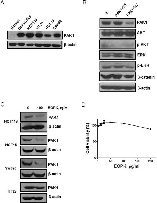
Background: The essential oil of Pinus koraiensis (EOPK) is biologically active compound obtained from the leaves of P. koraiensis. The goal of this study was to investigate the anti-cancer mechanism of EOPK in HCT116 colorectal cancer cells.

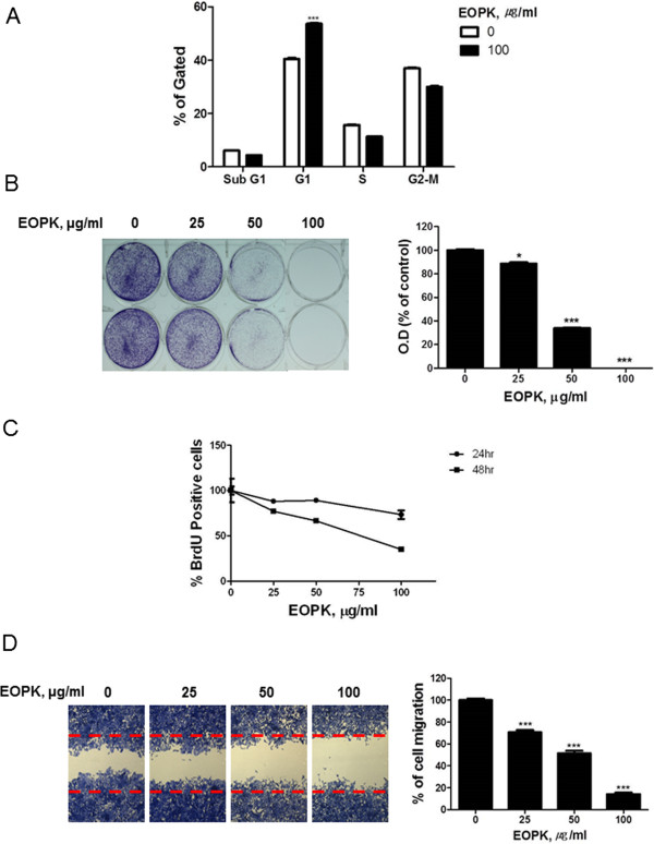
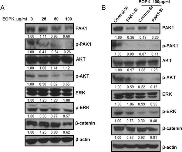
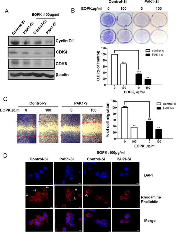
Methods: HCT116 cell proliferation was assessed by conducting crystal violet and BrdU assays. To assess the effects of EOPK on cell migration, we performed a wound-healing assay. Further, the contribution of PAK1 to EOPK-induced AKT and extracellular signal-regulated kinase (ERK) suppression was assessed by siRNA-mediated PAK1 knockdown. Changes to the expression and phosphorylation of PAK1 and its effectors were determined by western blotting, and changes to the actin cytoskeleton were determined by performing an immunofluorescence assay.

Results: EOPK significantly decreased HCT116 cell proliferation and migration, and induced G1 arrest without affecting normal cells. Additionally, EOPK suppressed the expression of PAK1, and decreased ERK and AKT phosphorylation in HCT116 cells. Finally, EOPK suppressed β-catenin, cyclin D1, and CDK4/6 expression.

Conclusions: Our studies indicate that EOPK significantly reduced proliferation and migration of colorectal cancer cells. Furthermore, EOPK suppressed PAK1 expression in a dose-dependent manner, and this suppression of PAK1 led to inhibition of ERK, AKT, and β-catenin activities. Our findings suggest that EOPK exerts its anticancer activity via the inhibition of PAK1 expression, suggesting it may be a potent chemotherapeutic agent for colorectal cancer.


Figures

Figure 1
Figure 1
PAK1 mediates MEK/ERK, PI3K/AKT, and Wnt/β-catenin in colon cancer cells. (A) Basal levels of PAK1 expression in colorectal cancer cell lines (human and murine) and normal colon cells extracted from mouse tissue were determined by Western blotting. (B) HCT116 cells were transfected with PAK1-siRNA1, PAK1-siRNA2, or control siRNA for 48 h. Cell lysates were prepared and subjected to Western blotting to determine the expression of PAK1, p-PAK1, AKT, p-AKT, ERK, p-ERK, β-catenin and β-actin. (C) Various human colorectal cancer cells were treated with EOPK (100 μg/ml) for 24 h. (D) Cytotoxicity of EOPK was analyzed by MTT assay in NIH-3 T3 cells as a normal cell control.
Figure 2
Figure 2
EOPK inhibits cell proliferation and migration in HCT116 colorectal cancer cells. (A) Cells were treated with EOPK (100 μg/ml) for 24 h. Cell cycle distribution was analyzed by flow cytometry. Bar graphs represent the percentage of sub-G1, G1, S, G2-M phase cells. Data represent mean ± SD of three independent experiments. *** p < 0.001 compared with untreated control. (B) HCT116 cells were treated with various concentrations of EOPK (25, 50, 100 μg/ml) and maintained for 5 days. The cells were resolved in 70% ethanol after washing with distilled water, and crystal violet absorbance was read using a microplate reader. Data represent mean ± SD of three independent experiments. * p < 0.05, ** p < 0.01 and *** p < 0.001 compared with control. (C) HCT116 cells were treated with EOPK for 24 h or 48 h and cell proliferation was measured by using a BrdU proliferation ELISA kit (Roche, Swiss). (D) Cells were treated with EOPK (25, 50, 100 μg/ml) for 24 h, and cell migration was assayed by wound healing assay. The number of cells migrating into the scratched area was photographed (×100) and calculated as a percentage of migration. Data are shown as the mean ± SD of three independent experiments. *** p < 0.001 compared with untreated control.
Figure 3
Figure 3
EOPK suppresses AKT and ERK phosphorylation and β-catenin expression via PAK1 inhibition in HCT116 colorectal cancer cells. (A) HCT116 cells were treated with various concentrations of EOPK (25, 50, 100 μg/ml). (B) HCT116 cells were transfected with PAK1-siRNA or a control siRNA for 48 h and incubated in the presence or absence of EOPK (100 μg/ml) for 24 h. For both experiments, cell lysates were prepared and subjected to Western blotting to determine the expression of PAK1, p-PAK1, AKT, p-AKT, ERK, p-ERK, β-catenin and β-actin. Band density of PAK1, p-PAK1, AKT, p-AKT, ERK, p-ERK, and β-catenin was quantified using Gelpro analyzer (Media Cybernetics, Bethesda, MD, USA).
Figure 4
Figure 4
PAK1 siRNA enhances the inhibitory effect of EOPK on the cell proliferation and migration. (A) HCT116 cells were transfected with PAK1-siRNA for 48 h and were incubated in the presence or absence of EOPK (100 μg/ml) for 24 h. Cell lysates were prepared and subjected to Western blotting to determine the expression of Cyclin D1, CDK4, CDK6 and β-actin. (B) HCT116 cells were transfected with PAK1-siRNA for 24 h and treated with EOPK (100 μg/ml) for 5 days. The cells were resolved in 70% ethanol after washing with distilled water, and crystal violet absorbance was read using a microplate reader. Data represent mean ± SD of three independent experiments. * p < 0.05, ** p < 0.01 and *** p < 0.001 compared with control. ### p < 0.001 compared with control-siRNA and PAK1-siRNA. (C) Cell migration was assessed by a wound healing assay. The number of cells migrated into the scratched area was calculated. Data are shown as the mean ± SD of three independent experiments. *** p < 0.001 compared with untreated control. ### p < 0.001 compared with control-siRNA and PAK1-siRNA. (D) Fixed cells were stained with rhodamine phalloidin and DAPI, and imaged using a DV Elite™ Imaging System (Deltavision, Applied Precision, Inc., USA). Arrow indicates lamellipodia. Arrow indicates lamellipodia.

References

    1. Sala-Vila A, Folkes J, Calder PC. The effect of three lipid emulsions differing in fatty acid composition on growth, apoptosis and cell cycle arrest in the HT-29 colorectal cancer cell line. Clin Nutr. 2010;29(4):519–524. doi: 10.1016/j.clnu.2009.11.004. - DOI - PubMed
    1. Calvert PM, Frucht H. The genetics of colorectal cancer. Ann Intern Med. 2002;137(7):603–612. - PubMed
    1. Yuen ST, Davies H, Chan TL, Ho JW, Bignell GR, Cox C, Stephens P, Edkins S, Tsui WW, Chan AS, Futreal PA, Stratton MR, Wooster R, Leung SY. Similarity of the phenotypic patterns associated with BRAF and KRAS mutations in colorectal neoplasia. Cancer Res. 2002;62(22):6451–6455. - PubMed
    1. Baldus SE, Schaefer KL, Engers R, Hartleb D, Stoecklein NH, Gabbert HE. Prevalence and heterogeneity of KRAS, BRAF, and PIK3CA mutations in primary colorectal adenocarcinomas and their corresponding metastases. Clin Cancer Res. 2010;16(3):790–799. doi: 10.1158/1078-0432.CCR-09-2446. - DOI - PubMed
    1. Fransen K, Klintenas M, Osterstrom A, Dimberg J, Monstein HJ, Soderkvist P. Mutation analysis of the BRAF, ARAF and RAF-1 genes in human colorectal adenocarcinomas. Carcinogenesis. 2004;25(4):527–533. doi: 10.1093/carcin/bgh049. - DOI - PubMed
Pre-publication history
    1. The pre-publication history for this paper can be accessed here:http://www.biomedcentral.com/1472-6882/14/275/prepub

Publication types