. 2014 Sep 12;289(37):25783-96.
doi: 10.1074/jbc.M114.577353. Epub 2014 Jul 29.

Coronavirus Nsp10, a critical co-factor for activation of multiple replicative enzymes

Affiliations

Coronavirus Nsp10, a critical co-factor for activation of multiple replicative enzymes

Mickaël Bouvet et al. J Biol Chem. .

Abstract

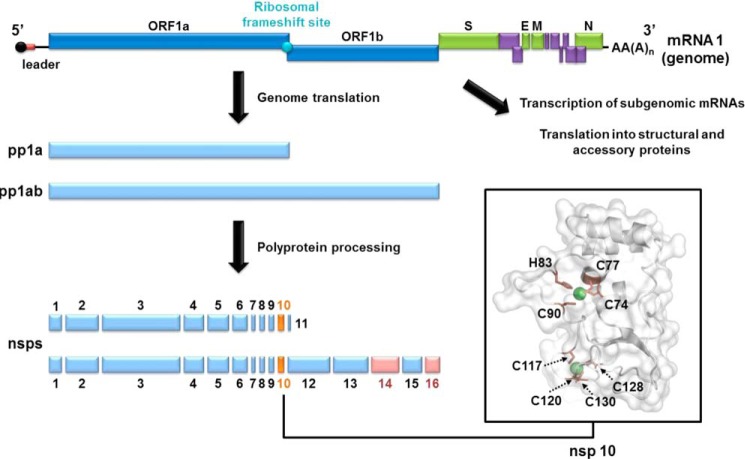
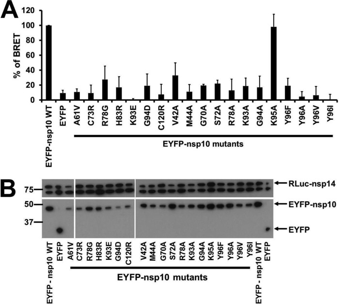
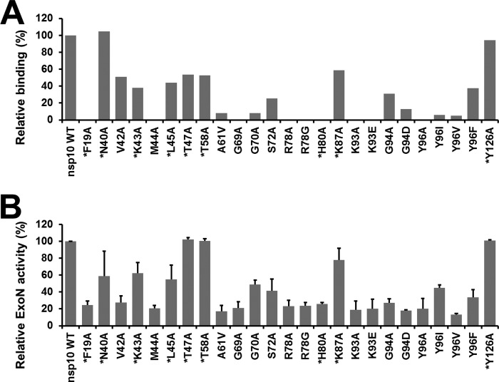
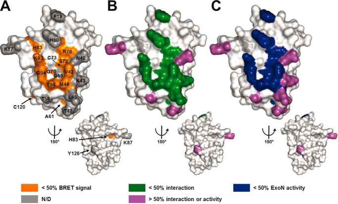
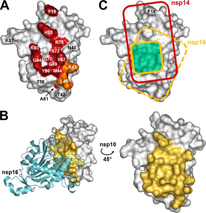
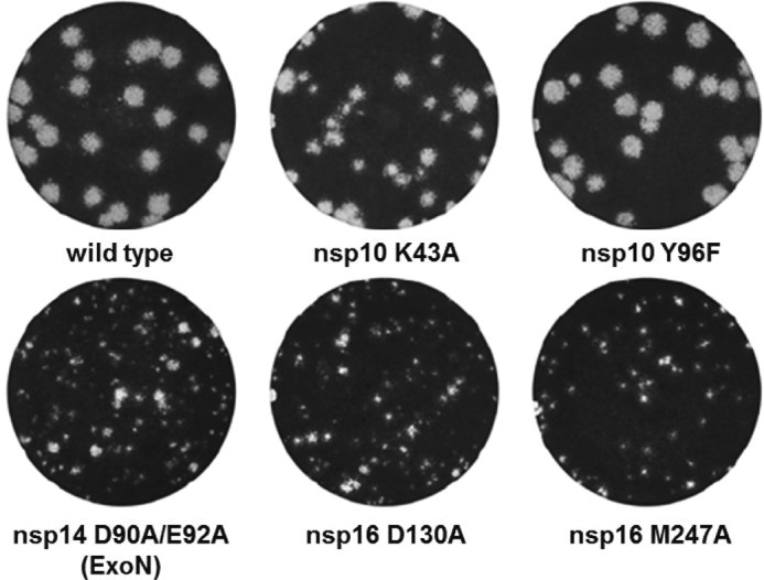
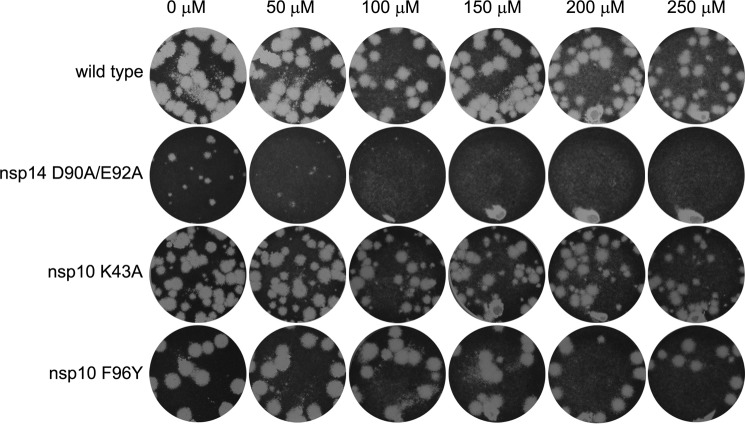
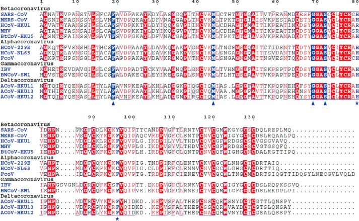
The RNA-synthesizing machinery of the severe acute respiratory syndrome Coronavirus (SARS-CoV) is composed of 16 non-structural proteins (nsp1-16) encoded by ORF1a/1b. The 148-amino acid nsp10 subunit contains two zinc fingers and is known to interact with both nsp14 and nsp16, stimulating their respective 3'-5' exoribonuclease and 2'-O-methyltransferase activities. Using alanine-scanning mutagenesis, in cellulo bioluminescence resonance energy transfer experiments, and in vitro pulldown assays, we have now identified the key residues on the nsp10 surface that interact with nsp14. The functional consequences of mutations introduced at these positions were first evaluated biochemically by monitoring nsp14 exoribonuclease activity. Disruption of the nsp10-nsp14 interaction abrogated the nsp10-driven activation of the nsp14 exoribonuclease. We further showed that the nsp10 surface interacting with nsp14 overlaps with the surface involved in the nsp10-mediated activation of nsp16 2'-O-methyltransferase activity, suggesting that nsp10 is a major regulator of SARS-CoV replicase function. In line with this notion, reverse genetics experiments supported an essential role of the nsp10 surface that interacts with nsp14 in SARS-CoV replication, as several mutations that abolished the interaction in vitro yielded a replication-negative viral phenotype. In contrast, mutants in which the nsp10-nsp16 interaction was disturbed proved to be crippled but viable. These experiments imply that the nsp10 surface that interacts with nsp14 and nsp16 and possibly other subunits of the viral replication complex may be a target for the development of antiviral compounds against pathogenic coronaviruses.

Keywords: Genetics; Protein-Protein Interaction; RNA Methyltransferase; RNA Virus; Viral Replication; Viral Transcription.


Figures

FIGURE 1.
FIGURE 1.
Schematic representation of SARS-CoV genome and nsp10 structure. Nsp10 (orange) and nsp14/16 (pink) are highlighted. The structural (green) and accessory (purple) protein genes are expressed from a nested set of subgenomic mRNAs. The schematic of the nsp10 structure (PDB code 2FYG) was generated using PyMOL software. Zinc ions are shown as green spheres, and residues forming the two nsp10 zinc fingers are labeled and depicted as brown sticks.
FIGURE 2.
FIGURE 2.
BRET characterization of the interaction of wild-type and mutant nsp10 with nsp14 in mammalian cells. A, BRET interaction assays were performed in HEK 293T cells after co-transfection of plasmids expressing EYFP-nsp10 mutants with an RLuc-nsp14 expressing plasmids. The experiments were performed three times, and the relative interaction of each mutant is calculated compared with the interaction of wild-type nsp10 with nsp14 (which was taken to be 100%). The BRET signals were further normalized according to the fluorescence signal measured for nsp10-EYFP mutants compared with wild-type control. B, Western blot analysis confirming similar levels of protein expression for the two interaction partners. Levels of RLuc-nsp14 and EYFP-nsp10 were determined using anti-luciferase and anti-GFP antibodies, respectively. The anti-luciferase antibody also recognized an ∼70-kDa host cell protein, which could conveniently serve as a loading control for the cell lysates.
FIGURE 3.
FIGURE 3.
Effect of nsp10 mutations on nsp10-nsp14 complex formation and on nsp14-ExoN activity in vitro. A, bar graph showing the relative nsp14 binding to each nsp10 mutant, as measured by in vitro pulldown assays. Nsp10 was purified by affinity chromatography and analyzed using capillary electrophoresis. The amount of nsp14 interacting with nsp10 was then quantified and normalized using nsp10. The binding activities were compared with the interaction of wild-type nsp10 with nsp14, which was arbitrarily set to 100%. B, relative nsp14 ExoN activities in the presence of a panel of nsp10 mutants. The ExoN activity obtained in the presence of wild-type nsp10 was arbitrarily set to 100%. Each experiment was repeated two times independently. Residues surrounding the nsp10 surface (as defined by BRET assay) that were newly included at this stage of the study are marked with an asterisk.
FIGURE 4.
FIGURE 4.
Three-dimensional structure of nsp10 highlighting residues involved in the interaction with nsp14. SARS-CoV nsp10 (PDB code 2FYG (37)) is depicted in white as a surface representation. A, residues that were found to be involved in interaction between nsp10 and nsp14 by the in vivo BRET assay (>50% effect) are colored in orange. B, residues that were found to be involved in the nsp10-nsp14 interaction according to the in vitro binding capacity (pulldown) assays (>50% effect) are colored in green. C, residues that were found to be involved in the nsp10 and nsp14 interaction on the basis of ExoN activity (>50% reduction) are colored in dark blue. Residues that could be mutated without significantly altering the nsp10-nsp14 interaction or the ExoN activity (<50% effect) are depicted in purple. Residues not tested in BRET experiments compared with other assays are displayed in gray (N/D). All figures were generated using PyMOL.
FIGURE 5.
FIGURE 5.
Comparison of the nsp14 and nsp16 interaction domains on the nsp10 surface. A, schematic representation of nsp10 residues that are engaged in the nsp10-nsp14 interaction. SARS-CoV nsp10 (PDB code 2FYG (37)) is shown as a white surface representation. Residues that significantly affect the nsp10-nsp14 interaction when mutated are colored in red (>50% decrease of BRET values, binding affinity, and ExoN activity). Residues Lys-43 and Leu-45, which impair the nsp10-nsp14 interaction with a smaller effect when mutated (both binding affinity and ExoN activity ∼50% of wild type), are shown in orange. B, representation of nsp10 residues involved in the nsp10-nsp16 interaction in the structure of the complex (PDB code 2XYQ (40)). Nsp16 is shown as a schematic representation, colored in cyan. Nsp10 residues that are present within a 5 Å radius of nsp16 are depicted in yellow. C, schematic representation of nsp10 functional interacting surface with nsp14 (red rectangle) and nsp16 (dark yellow square, based on Lugari et al. (41)). The structural nsp10-nsp16 interaction surface is depicted as a dashed yellow rectangle on nsp10. The overlapping functional interaction surface is depicted in pale green. All figures were generated using PyMOL.
FIGURE 6.
FIGURE 6.
Plaque morphology of viable SARS-CoV nsp10, nsp14, and nsp16 mutants. Small-plaque phenotypes were observed for mutants nsp14-D90A/E92A, nsp16-D130A, and nsp16-M247A, an intermediate plaque size for mutant nsp10-K43A, whereas the plaque size of mutant nsp10-Y96F was similar to that of the wild-type control.
FIGURE 7.
FIGURE 7.
Lack of 5′-fluorouracil sensitivity of SARS-CoV mutants nsp10-K43A and nsp10-Y96F suggests they do not exhibit a mutator phenotype. Plaque reduction assays were performed in the presence of increasing concentrations of 5-fluorouracil. Each well was infected with the same amount of virus, after which cell layers were overlaid with a semi-solid medium containing the indicated increasing concentrations of 5FU. Cell layers were incubated for 3 days, fixed, and stained to reveal plaque formation. Like the wild-type virus, nsp10 mutants K43A and Y96F were insensitive to 5FU up to a dose of 250 μm. On the other hand, plaque size and number for mutant nsp14-D90A/E92A (the ExoN knock-out mutant known to exhibit a mutator phenotype) were strongly reduced even at the lowest 5FU concentration tested.
FIGURE 8.
FIGURE 8.
Conservation of the nsp10 sequence across the Coronaviridae subfamily. Alignment of nsp10 sequences from a representative set of Coronavirinae subfamily viruses including members of each of the four genera (Alpha-, Beta-, Gamma-, and Deltacoronavirus). Residues that are conserved in all sequences and whose replacement is lethal to SARS-CoV and MHV (Phe-19, Met-44, Gly-69, and Ser-72) are boxed in blue and indicated by triangles. Residues that are conserved in >80% of the sequences and whose replacement is lethal to SARS-CoV (His-80 and Tyr-96) are labeled in blue and indicated by asterisks. Sequences were aligned using the ESPript program (67). National Center for Biotechnology Information (NCBI) accession numbers for replicase polyprotein sequences including nsp10 are: SARS-CoV, AY345988; MERS-CoV, JX869059; HCoV-HKU1, AY884001; MHV, AY700211; BtCoV-HKU5, bat Coronavirus HKU5–1 (EF065509); HCoV-229E, NC_002645; HCoV-NL63, DQ445911; FcoV, feline Coronavirus (DQ010921); IBV, avian infectious bronchitis virus (NC_001451); BWCoV-SW1, beluga whale Coronavirus SW1 (EU111742); ACoV-HKU11, bulbul Coronavirus HKU11-796 (FJ376620); ACoV-HKU13, munia Coronavirus HKU13-3514 (NC_011550); ACoV-HKU12, thrush Coronavirus HKU12-600 (NC_011549).

Similar articles

Cited by

References

    1. Gorbalenya A. E., Enjuanes L., Ziebuhr J., Snijder E. J. (2006) Nidovirales: evolving the largest RNA virus genome. Virus Res. 117, 17–37 - PMC - PubMed
    1. Nga P. T., Parquet Mdel C., Lauber C., Parida M., Nabeshima T., Yu F., Thuy N. T., Inoue S., Ito T., Okamoto K., Ichinose A., Snijder E. J., Morita K., Gorbalenya A. E. (2011) Discovery of the first insect Nidovirus, a missing evolutionary link in the emergence of the largest RNA virus genomes. PLoS Pathog. 7, e1002215. - PMC - PubMed
    1. Peiris J. S., Guan Y., Yuen K. Y. (2004) Severe acute respiratory syndrome. Nat. Med. 10, S88–S97 - PMC - PubMed
    1. Smith I., Wang L. F. (2013) Bats and their virome: an important source of emerging viruses capable of infecting humans. Curr. Opin. Virol. 3, 84–91 - PMC - PubMed
    1. Corman V. M., Kallies R., Philipps H., Göpner G., Müller M. A., Eckerle I., Brünink S., Drosten C., Drexler J. F. (2014) Characterization of a novel Betacoronavirus related to middle East respiratory syndrome Coronavirus in European hedgehogs. J. Virol. 88, 717–724 - PMC - PubMed

Publication types

MeSH terms

Substances