

doi: 10.3201/eid2008.140663.
Isolation of MERS coronavirus from a dromedary camel, Qatar, 2014
- PMID: 25075761
- PMCID: PMC4111206
- DOI: 10.3201/eid2008.140663
Item in Clipboard
Isolation of MERS coronavirus from a dromedary camel, Qatar, 2014
Emerg Infect Dis.
2014 Aug.
Abstract
We obtained the full genome of Middle East respiratory syndrome coronavirus (MERS-CoV) from a camel in Qatar. This virus is highly similar to the human England/Qatar 1 virus isolated in 2012. The MERS-CoV from the camel efficiently replicated in human cells, providing further evidence for the zoonotic potential of MERS-CoV from camels.
Keywords: MERS; Qatar; camel; coronavirus; viruses.
Figures
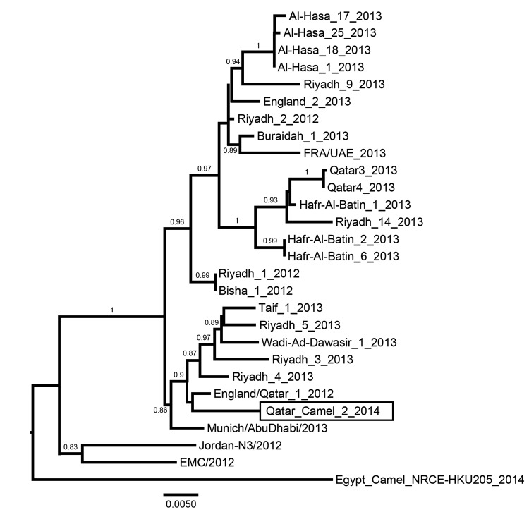
Phylogenetic analysis of Middle East respiratory syndrome coronaviruses (MERS-CoVs). Genome sequences of representative isolates were aligned by using ClustalW, and a phylogenetic tree was constructed by using the PhyML method in Seaview 4 (all 3 software packages can be found at http://pbil.univ-lyon1.fr/software/seaview ) and was visualized in FigTree version 1.3.1 (http://tree.bio.ed.ac.uk/software/figtree/ ). Values at branches show the result of the approximate likelihood ratio; values <0.70 are not shown. The MERS-CoV isolated from a dromedary camel in Qatar in 2014 is depicted in a rectangle. Scale bar indicates nucleotide substitutions per site.
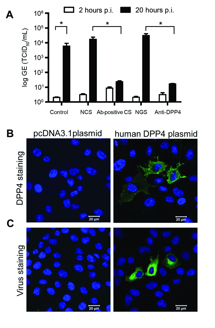
Middle East respiratory syndrome coronavirus (MERS-CoV) from camel replicates in human hepatoma (Huh-7) cells and uses human DPP4 as entry receptor. Huh-7 cells were inoculated with camel MERS-CoV and left for 1 h. Next, cells were washed twice, and supernatant was collected at 2 h (open bars) and 20 h (closed bars) before being tested for MERS-CoV RNA by using a TaqMan assay. We analyzed control camel MERS-CoV–infected cells, cells inoculated with camel MERS-CoV in the presence of normal camel serum (NCS), MERS-CoV–antibody positive camel serum (Ab-positive CS), normal goat serum (NGS), and anti-DPP4 polyclonal antibody–treated cells. Results are expressed as genome equivalents (GE), 50% tissue culture infective dose (TCID50/mL) (A). MDCK cells transfected with plasmid-encoding human DPP4 or a control plasmid (pcDNA) were stained with polyclonal antibody against human DPP4 (B) or inoculated with camel MERS-CoV and fixed 20 h after inoculation (p.i.) and stained for viral antigen (C).
References
-
- Hemida MG, Perera R, Wang P, Alhammadi MA, Siu LY, Poon LL, et al. Middle East respiratory syndrome (MERS) coronavirus seroprevalence in domestic livestock in Saudi Arabia, 2010 to 2013. Euro Surveill. 2013;18:20659 . - PubMed
Publication types
MeSH terms
Substances
Associated data
- Actions
LinkOut - more resources
Full Text Sources
Other Literature Sources
