. 2015 Jan;87(1):151-61.
doi: 10.1038/ki.2014.268. Epub 2014 Jul 30.

Ribonucleases 6 and 7 have antimicrobial function in the human and murine urinary tract

Affiliations

Ribonucleases 6 and 7 have antimicrobial function in the human and murine urinary tract

Brian Becknell et al. Kidney Int. 2015 Jan.

Abstract

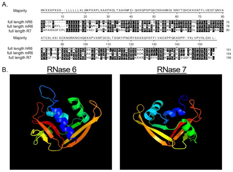
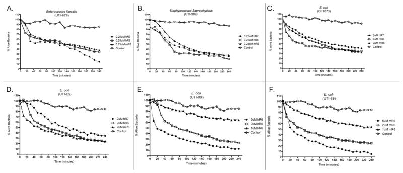
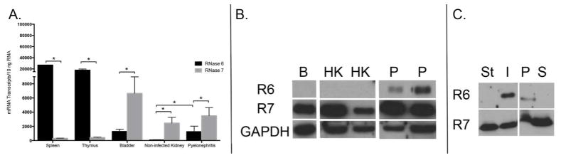
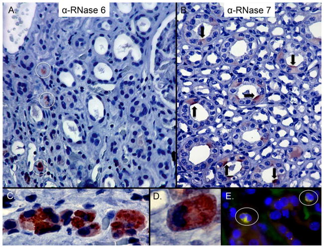
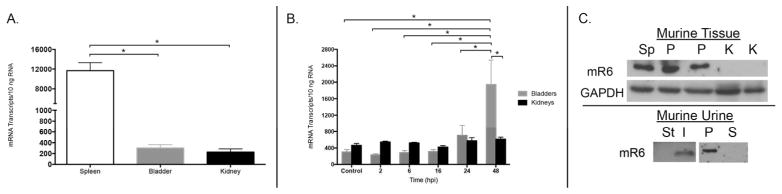
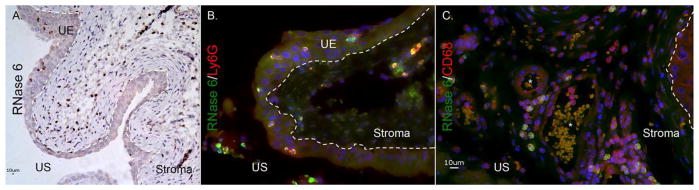
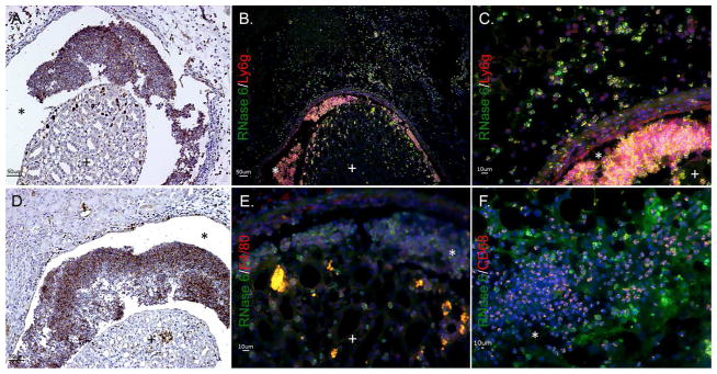
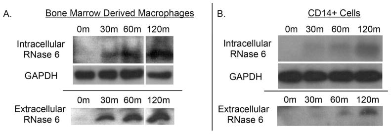
Recent evidence suggests antimicrobial peptides protect the urinary tract from infection. Ribonuclease 7 (RNase 7), a member of the RNase A superfamily, is a potent epithelial-derived protein that maintains human urinary tract sterility. RNase 7 expression is restricted to primates, limiting evaluation of its antimicrobial activity in vivo. Here we identified ribonuclease 6 (RNase 6) as the RNase A superfamily member present in humans and mice that is most conserved at the amino acid level relative to RNase 7. Like RNase 7, recombinant human and murine RNase 6 has potent antimicrobial activity against uropathogens. Quantitative real-time PCR and immunoblot analysis indicate that RNase 6 mRNA and protein are upregulated in the human and murine urinary tract during infection. Immunostaining located RNase 6 to resident and infiltrating monocytes, macrophages, and neutrophils. Uropathogenic E. coli induces RNase 6 peptide expression in human CD14(+) monocytes and murine bone marrow-derived macrophages. Thus, RNase 6 is an inducible, myeloid-derived protein with markedly different expression from the epithelial-derived RNase 7 but with equally potent antimicrobial activity. Our studies suggest RNase 6 serves as an evolutionarily conserved antimicrobial peptide that participates in the maintenance of urinary tract sterility.


Conflict of interest statement

DISCLOSURE

None of the authors declares a financial interest in the content of this manuscript.

Figures

Figure 1
Figure 1. Amino acid homology and predicted structural similarities between mouse RNase6, human RNase 6, and RNase 7
(A) Alignment of full length proteins by Clustal W method. Residues matching the consensus (“majority”) are shaded. (B) Predicted solution structure of human RNase 6 and RNase 7.,
Figure 2
Figure 2. RNase 6 and RNase 7 rapidly kill uropathogenic bacteria at micromolar concentrations
Uropathogenic E. faecalis (A), S. saprophyticus (B), and E. coli strains CFT073 and UTI89 (C,D) were stained using a 1:1 mixture of SYTO®9, which labels live bacteria, and propidium iodide, which labels killed bacteria. Bacteria were incubated with fixed concentrations of recombinant murine RNase 6, human RNase 6, or RNase 7. (E/F) E. coli strain UTI89 was stained as outlined above and incubated with increasing concentrations of recombinant human or mouse RNase 6. Untreated bacteria served as the control. Bacterial viability over time was analyzed integrating fluorescent changes in SYTO®9 dye and propidium iodide dye. Values are the average of three replicates.
Figure 3
Figure 3. RNase 6 and RNase 7 mRNA and peptide expression in the human urinary tract
(A) Quantification of human RNASE6 and RNASE7 mRNA in non-infected tissues and pyelonephritis kidney samples by qRT-PCR. (B) Western immunoblot analysis of human RNase 6 (R6) and RNase 7 (R7) protein in bladder (B), non-infected kidney (HK) and pyelonephritis (P) lysates. GAPDH is included as a loading control. (C) Immunoblot analysis of human RNase 6 (R6) and RNase 7 (R7) in sterile (St) or UPEC infected (I) human urine (left). Infected human urine was subjected to centrifugation and immunoblot analysis was repeated using the cellular pellet (P) and supernatant (S) fractions (right).
Figure 4
Figure 4. Expression of RNase 6 and RNase 7 in the human kidney
(A) Immunohistochemistry demonstrates RNase 6 production (brown/circles) in isolated, interstitial leukocytes in the renal parenchyma (B) and RNase 7 production (brown/arrows) in isolated renal tubules. Epithelial expression of RNase 6 was not routinely detected. Original Magnification 20×. (C/D) High-power magnification identifies RNase 6 production by granulocytes with greatest expression in the cytoplasmic granules. Original magnification 40× and 100×, respectively. (E) Human pyelonephritis samples were labeled with RNase 6 (green), CD68 (red), and nuclei (blue). RNase 6 expression was detected in CD68-positive cells. Original magnification 40×.
Figure 5
Figure 5. Expression of RNase 6 and RNase 7 in the human lower urinary tract
Immunohistochemistry demonstrates RNase 6 production (brown/circles) in isolated urothelial and submucosal leukocytes in the human ureter (A) whereas RNase 7 (brown/arrows) is produced by the apical uroepithelium (UE) of the lower urinary tract (B). Original magnification 40×. (C/D/E) Immunohistochemistry demonstrates RNase 6 production (red/circles) in intravascular leukocytes of human bladder. Original Magnification 100×.
Figure 6
Figure 6. RNase 6 mRNA and RNase 6 peptide expression in the murine urinary tract
(A) Quantification of mouse Rnase6 mRNA in sterile tissues by qRT-PCR. Mean ± S.E.M. are shown from 4 bladders and kidneys. (B) Rnase6 mRNA expression over time in bladder and kidneys of UPEC infected mice. Time in hours post infection (hpi) is indicated on the X-axis. Mean ± S.E.M. are shown from 4 bladders and kidneys at each time point. (C, top) Western immunoblot of RNase 6 protein in mouse spleen (Sp), non-infected kidney (K), and pyelonephritis (P) kidney lysates. GADPH is included as a loading control. (C, bottom) Western immunoblot analysis of mouse RNase 6 peptide in sterile (St) and UPEC infected (I) urine samples (left). Infected mouse urine was subject to centrifugation and immunoblot analysis was repeated using the cellular pellet (P) and supernatant (S) fractions (right).
Figure 7
Figure 7. RNase 6 peptide localization in the lower murine urinary tract
(A) Immunohistochemistry demonstrates RNase 6 production (brown) in isolated uroepithelial and stromal leukocytes in UPEC-infected mouse bladder. Original magnification 20×. (B/C) Infected mouse bladders labeled for RNase 6 (green), the neutrophil marker Ly6g (red) or the monocyte marker CD68 (red), and nuclei (blue). RNase 6 expression was detected in Ly6g and CD68-positive cells. Original magnification 40×. The dashed line demarcates the uroepithelium (UE) and the stromal layers of the bladder. US: urinary space.
Figure 8
Figure 8. RNase 6 peptide localization in the murine kidney
(A/D) Immunohistochemistry identifies RNase 6 production (brown) in isolated leukocytes in UPEC infected murine kidneys. Original Magnification 20×. (B/C) UPEC infected mouse kidneys were labeled with RNase 6 (green), the neutrophil marker Ly6g (red), and nuclei (blue). RNase 6 was detected in Ly6G positive neutrophils. (E/F) UPEC infected mouse kidneys were labeled with RNase 6 (green), the monocyte/macrophage markers F4/80 or CD68 (red), and nuclei (blue). RNase 6 was detected in CD68 positive cells. Original Magnification 40×. (+) identifies the renal parenchyma and (*) identifies the urinary space.
Figure 9
Figure 9. RNase 6 peptide production by monocyte/macrophages following bacterial exposure
(A) Murine bone marrow derived macrophages and (B) human CD14(+) monocytes, were incubated with UPEC strain CFT073 for the indicated times and RNase 6 peptide was detected in cellular pellets and supernatants by Western immunoblot analysis.

References

    1. Foxman B, Barlow R, D’Arcy H, Gillespie B, Sobel JD. Urinary tract infection: self-reported incidence and associated costs. Ann Epidemiol. 2000;10:509–15. - PubMed
    1. Spencer JD, Schwaderer AL, Becknell B, Watson J, Hains DS. The innate immune response during urinary tract infection and pyelonephritis. Pediatr Nephrol. 2013 - PMC - PubMed
    1. Zasloff M. Antimicrobial peptides, innate immunity, and the normally sterile urinary tract. J Am Soc Nephrol. 2007;18:2810–6. - PubMed
    1. Yeaman MR, Yount NY. Mechanisms of antimicrobial peptide action and resistance. Pharmacol Rev. 2003;55:27–55. - PubMed
    1. Spencer JD, Schwaderer AL, Dirosario JD, et al. Ribonuclease 7 is a potent antimicrobial peptide within the human urinary tract. Kidney Int. 2011;80:174–80. - PubMed

Publication types