

Orbital granulomatosis with polyangiitis (Wegener granulomatosis): clinical and pathologic findings
- PMID: 25076302
- PMCID: PMC4140401
- DOI: 10.5858/arpa.2013-0006-RS
Orbital granulomatosis with polyangiitis (Wegener granulomatosis): clinical and pathologic findings
Abstract
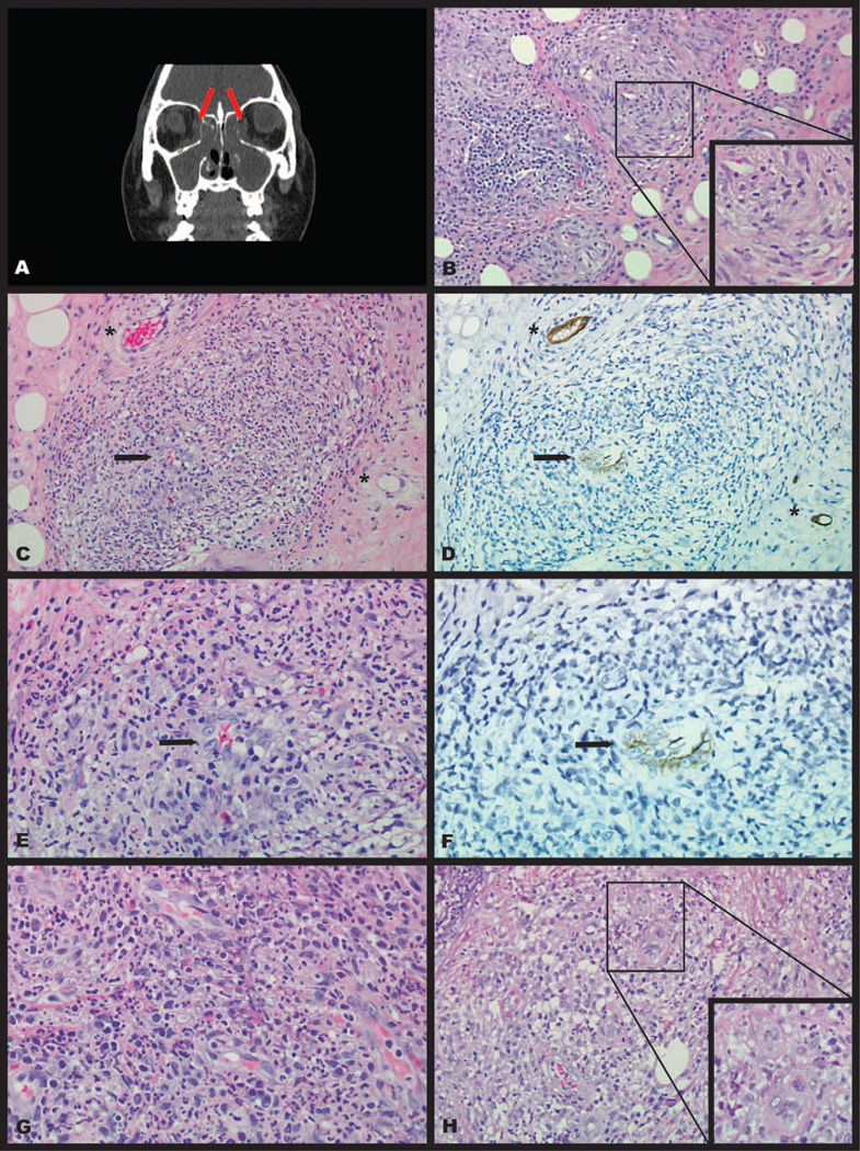
The pathology of granulomatosis with polyangiitis (GPA), formerly Wegener granulomatosis, typically features a granulomatous and sometimes necrotizing vasculitis targeting the respiratory tract and kidneys. However, orbital involvement occurs in up to 60% of patients and is frequently the first or only clinical presentation in patients with systemic or limited forms of GPA. Orbital GPA can cause significant morbidity and potentially lead to complete loss of vision and permanent facial deformity. Fortunately, GPA is highly responsive to medical treatment with corticosteroids combined with cyclophosphamide or, more recently, rituximab. Therefore, it is imperative for this disease to be accurately diagnosed on orbital biopsy and distinguished from other histologically similar orbital lesions. Herein, we review the clinical and pathologic findings of orbital GPA, focusing on the differentiation of this disease from other inflammatory orbital lesions.
Figures
References
-
- Falk RJ, Gross WL, Guillevin L, et al. Granulomatosis with polyangiitis (Wegener’s): an alternative name for Wegener’s granulomatosis. Ann Rheum Dis. 2011;70(4):704. - PubMed
-
- Fahey JL, Leonard E, Churg J, Godman G. Wegener’s granulomatosis. Am J Med. 1954;17(2):168–179. - PubMed
-
- Leavitt RY, Fauci AS, Bloch DA, et al. The American College of Rheumatology 1990 criteria for the classification of Wegener’s granulomatosis. Arthritis Rheum. 1990;33(8):1101–1107. - PubMed
-
- Savige J, Dimech W, Fritzler M, et al. Addendum to the International Consensus Statement on testing and reporting of antineutrophil cytoplasmic antibodies: quality control guidelines, comments, and recommendations for testing in other autoimmune diseases. Am J Clin Pathol. 2003;120(3):312–318. - PubMed
-
- Breda L, Nozzi M, De Sanctis S, Chiarelli F. Laboratory tests in the diagnosis and follow-up of pediatric rheumatic diseases: an update. Semin Arthritis Rheum. 2010;40(1):53–72. - PubMed
Publication types
MeSH terms
Substances
Grants and funding
LinkOut - more resources
Full Text Sources
Other Literature Sources
Medical