. 2014 Mar 21:9034:90342V.
doi: 10.1117/12.2043260.

An artifact-robust, shape library-based algorithm for automatic segmentation of inner ear anatomy in post-cochlear-implantation CT

Affiliations

An artifact-robust, shape library-based algorithm for automatic segmentation of inner ear anatomy in post-cochlear-implantation CT

Fitsum A Reda et al. Proc SPIE Int Soc Opt Eng. .

Abstract

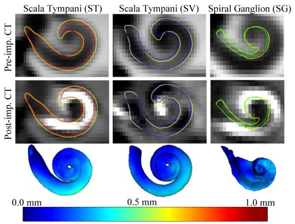
A cochlear implant (CI) is a device that restores hearing using an electrode array that is surgically placed in the cochlea. After implantation, the CI is programmed to attempt to optimize hearing outcome. Currently, we are testing an image-guided CI programming (IGCIP) technique we recently developed that relies on knowledge of relative position of intracochlear anatomy to implanted electrodes. IGCIP is enabled by a number of algorithms we developed that permit determining the positions of electrodes relative to intra-cochlear anatomy using a pre- and a post-implantation CT. One issue with this technique is that it cannot be used for many subjects for whom a pre-implantation CT was not acquired. Pre-implantation CT has been necessary because it is difficult to localize the intra-cochlear structures in post-implantation CTs alone due to the image artifacts that obscure the cochlea. In this work, we present an algorithm for automatically segmenting intra-cochlear anatomy in post-implantation CTs. Our approach is to first identify the labyrinth and then use its position as a landmark to localize the intra-cochlea anatomy. Specifically, we identify the labyrinth by first approximately estimating its position by mapping a labyrinth surface of another subject that is selected from a library of such surfaces and then refining this estimate by a standard shape model-based segmentation method. We tested our approach on 10 ears and achieved overall mean and maximum errors of 0.209 and 0.98 mm, respectively. This result suggests that our approach is accurate enough for developing IGCIP strategies based solely on post-implantation CTs.

Keywords: CI programming; Cochlear implant (CI) surgery; intra-cochlear anatomy; registration; segmentation.


Figures

Figure 1
Figure 1
Shown in (a) and (b) are surfaces of the ST (red), the SV (blue), and the SG (green). In (b), a surface model of a CI electrode array inserted into the ST is shown. Panel (c) shows surfaces of the AR (green), the ST (transparent red), and the SV (transparent blue). Panel (d) shows contours of the ST (red), the SG (green), and the electrodes (purple) in the coronal view of a pre-implantation CT and a corresponding post-implantation CT. Shown in (e) are contours of the SV (blue) in the coronal view of a pre-implantation CT and a corresponding post-implantation CT.
Figure 2
Figure 2
Shown in (a) are surfaces of a labyrinth (transparent orange) and of the intra-cochlear anatomy (ST (transparent red), SV (transparent blue), and SG (transparent green)). Panel (b) shows the same structures in a different orientation. In (c) the set of points that represent the external wall of cochlea and that are used to fit the SOI model to the labyrinth model is shown on the surface of the labyrinth. In (d) and (e) the same set of points is shown on the SOI surfaces. Panel (f) shows a labyrinth surface with near points in yellow and far points in purple.
Figure 3
Figure 3
Image registration process.
Figure 4
Figure 4
Segmentation refinement process.
Figure 5
Figure 5
Surfaces of intra-cochlear structures colormapped with segmentation errors viewed on the coronal plane (top row) and sagittal plane (bottom row).
Figure 6
Figure 6
Results for a case with average error close to the overall average error. The contours shown are the ST (left panel), SV (middle panel), and SG (right panel). Contours for gold-standard ST (red), SV (blue), SG (green) surfaces and contours for automatic surfaces
Figure 7
Figure 7
Surface of the active region colormapped with segmentation errors for each testing ear.

References

    1. Noble JH, Labadie RF, Gifford RH, Dawant BM. Image-guidance enables new methods for customizing cochlear implant stimulation strategies. Neural Systems and Rehabilitation Engineering. 2013 Sept.21(5):820–829. IEEE Transactions on. - PMC - PubMed
    1. Noble JH, Labadie RF, Majdani O, Dawant BM. Automatic Segmentation of Intracochlear Anatomy in Conventional CT. Biomedical Engineering. 2011 Sept.58(9):2625–2632. IEEE Transactions on. - PMC - PubMed
    1. Noble Jack H., Schuman Theodore A., Wright Charles G., Labadie Robert F., Dawant Benoit M. Automatic identification of cochlear implant electrode arrays for post-operative assessment. Proc. SPIE 7962, Medical Imaging 2011: Image Processing; 796217. - PMC - PubMed
    1. Schuman TA, Noble JH, Wright CG, Wanna GB, Dawant B, Labadie RF. Anatomic Verification of a Novel, Non-rigid Registration Method for Precise Intrascalar Localization of Cochlear Implant Electrodes in Adult Human Temporal Bones Using Clinically-available Computerized Tomography. The Laryngoscope. 2010;120(11):2277–2283. - PMC - PubMed
    1. Wanna GB, Noble JH, McRackan TR, Dawant BM, Dietrich MS, Watkins LD, Rivas A, Schuman TA, Labadie RF. Assessment of electrode positions and audiological outcomes in bilateral cochlear implant patients. Otology & Neurotology. 2011;32(3):428–432. - PMC - PubMed