Review
. 2014 Jul 16:8:241.
doi: 10.3389/fnbeh.2014.00241. eCollection 2014.

Optogenetic insights on the relationship between anxiety-related behaviors and social deficits

Affiliations
Review

Optogenetic insights on the relationship between anxiety-related behaviors and social deficits

Stephen A Allsop et al. Front Behav Neurosci. .

Abstract

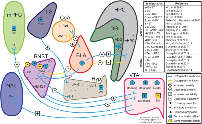
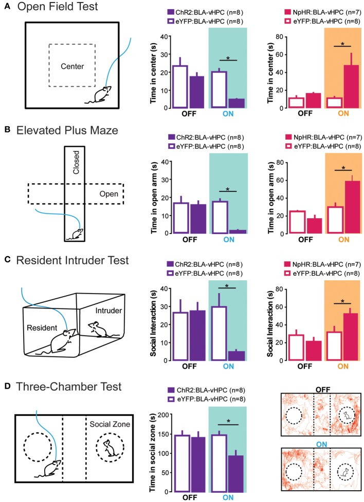
Many psychiatric illnesses are characterized by deficits in the social domain. For example, there is a high rate of co-morbidity between autism spectrum disorders and anxiety disorders. However, the common neural circuit mechanisms by which social deficits and other psychiatric disease states, such as anxiety, are co-expressed remains unclear. Here, we review optogenetic investigations of neural circuits in animal models of anxiety-related behaviors and social behaviors and discuss the important role of the amygdala in mediating aspects of these behaviors. In particular, we focus on recent evidence that projections from the basolateral amygdala (BLA) to the ventral hippocampus (vHPC) modulate anxiety-related behaviors and also alter social interaction. Understanding how this circuit influences both social behavior and anxiety may provide a mechanistic explanation for the pathogenesis of social anxiety disorder, as well as the prevalence of patients co-diagnosed with autism spectrum disorders and anxiety disorders. Furthermore, elucidating how circuits that modulate social behavior also mediate other complex emotional states will lead to a better understanding of the underlying mechanisms by which social deficits are expressed in psychiatric disease.

Keywords: anxiety; autism; basolateral amygdala; mouse models of affective disorders; optogenetics; social deficits; social interaction; ventral hippocampus.


Figures

Figure 1
Figure 1
Neural circuits implicated in anxiety and social function through optogenetic investigations. The ability to selectively manipulate distinct neuronal populations and projections with high temporal resolution is a significant advantage of optogenetic approaches. Several recent studies have used optogenetic strategies to establish causal relationships between specific neuronal projections and behaviors relevant to anxiety and sociability in non-stressed animals. Abbreviations: ad, anterodorsal subdivision of the bed nucleus of the stria terminalis; aMH, anterior hypothalamic area of the medial hypothalamus; CeA, central amygdala; Crfr2, type-2 corticotropin-releasing factor receptor; BLA, basolateral amygdala; BNST, bed nucleus of the stria terminalis; DA, dopaminergic neurons; DG, dentate gyrus; Drd1a, dopamine receptor 1a; E/I, manipulation of excitatory/inhibitory balance; GABA, GABAergic neurons; GCs, granule cells; HPC, hippocampus; Hyp, hypothalamus; LH, lateral hypothalamus; LS, lateral septum; mPFC, medial prefrontal cortex; NAc, nucleus accumbens; Orx, orexin neurons; ov, oval nucleus of the bed nucleus of the stria terminalis; pLH, posterior lateral hypothalamus; vBNST, ventral subdivision of the bed nucleus of the stria terminalis; vDG, ventral dentate gyrus; vHPC, ventral hippocampus; VTA, ventral tegmental area. (For review of optogenetic investigations into the neural circuitry involved in aggression and sexual behavior, we refer readers to Anderson, 2012).
Figure 2
Figure 2
Projections from the BLA to the vHPC bidirectionally modulate anxiety-related behaviors as well as social interaction. To perform all of the following experiments, channelrhodopsin-2 or an enhanced version of halorhodopsin was injected into the basolateral amygdala (BLA) and an optical fiber was placed above the ventral hippocampus (vHPC). This allowed selective optogenetic manipulation of BLA projections to the vHPC. (A) In the open field test, decreased time spent in the center was measured as a proxy for increased anxiety. Excitation of BLA-vHPC projections lead to an increase in anxiety-related behaviors while inhibition of BLA-vHPC projections lead to a decrease in anxiety-related behaviors. (B) In the elevated plus maze, decreased time in the open arms was measured as a proxy for increased anxiety. Excitation of BLA-vHPC projections lead to an increase in anxiety-related behaviors while inhibition of BLA-vHPC projections lead to a decrease in anxiety-related behaviors. (C) The resident intruder test was used to measure social interaction. The overall score of social interaction included body sniffing, anogenital sniffing, direct contact, and close following (<1 cm). Activation of BLA-vHPC projections led to a decrease in social interaction while inhibition of BLA-vHPC projections led to an increase in social interaction. (D) The three-chamber test was used as a test for sociability. Time spent in the social zone was measured as proxy for sociability. Activation of BLA-vHPC projections lead to a decrease in time spent in the social zone. (A–D) These experiments demonstrate that anxiety and social interaction can be modulated at the level of the neural circuit. Additionally activation of BLA-vHPC projections leads to increases in anxiety-related behaviors as well as decreases in social interaction while inhibition of the same circuit causes a decrease in anxiety-related behaviors as well as an increase in social interaction. Data are mean values and error bars represent SEM. For the Open Field Test and Elevated Plus Maze, the light-off epochs were pooled and averaged (One Way ANOVA with Bonferroni post test, *p < 0.01). For the Resident Intruder Test and the Three-Chamber Test statistics described in Felix-Ortiz and Tye (2014) (*p < 0.05).

References

    1. Adolphs R. (2003). Cognitive neuroscience of human social behaviour. Nat. Rev. Neurosci. 4, 165–178 10.1038/nrn1056 - DOI - PubMed
    1. Adolphs R. (2009). The social brain: neural basis of social knowledge. Annu. Rev. Psychol. 60, 693–716 10.1146/annurev.psych.60.110707.163514 - DOI - PMC - PubMed
    1. Adolphs R. (2010). What does the amygdala contribute to social cognition? Ann. N.Y. Acad. Sci. 1191, 42–61 10.1111/j.1749-6632.2010.05445.x - DOI - PMC - PubMed
    1. Adolphs R., Tranel D., Damasio H., Damasio A. (1994). Impaired recognition of emotion in facial expressions following bilateral damage to the human amygdala. Nature 372, 669–672 10.1038/372669a0 - DOI - PubMed
    1. Akimova E., Lanzenberger R., Kasper S. (2009). The serotonin-1A Receptor in anxiety disorders. Biol. Psychiatry 66, 627–635 10.1016/j.biopsych.2009.03.012 - DOI - PubMed

LinkOut - more resources