Review
. 2014 Jun 2:5:259.
doi: 10.3389/fimmu.2014.00259. eCollection 2014.

Rag defects and thymic stroma: lessons from animal models

Affiliations
Review

Rag defects and thymic stroma: lessons from animal models

Veronica Marrella et al. Front Immunol. .

Erratum in

  • Front Immunol. 2014;5:407. Grassi, Fabio [added]

Abstract

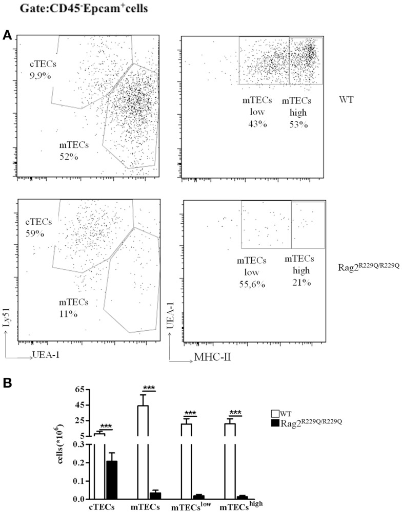
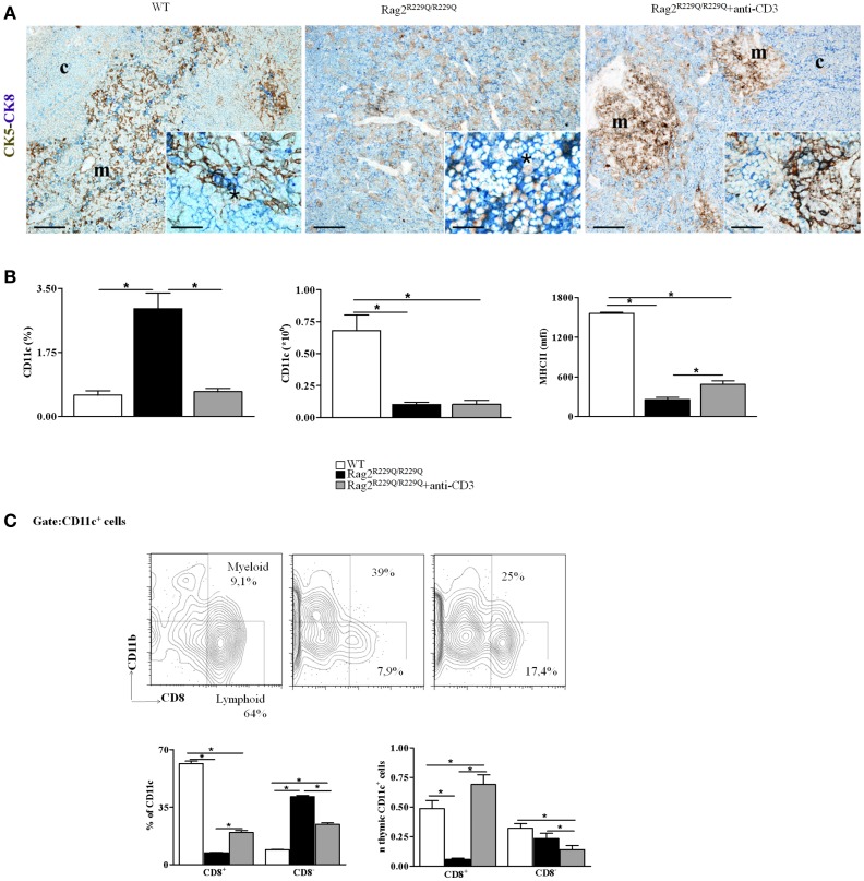
Thymocytes and thymic epithelial cells (TECs) cross-talk is essential to support T cell development and preserve thymic architecture and maturation of TECs and Foxp3(+) natural regulatory T cells. Accordingly, disruption of thymic lymphostromal cross-talk may have major implications on the thymic mechanisms that govern T cell tolerance. Several genetic defects have been described in humans that affect early stages of T cell development [leading to severe combined immune deficiency (SCID)] or late stages in thymocyte maturation (resulting in combined immunodeficiency). Hypomorphic mutations in SCID-causing genes may allow for generation of a limited pool of T lymphocytes with a restricted repertoire. These conditions are often associated with infiltration of peripheral tissues by activated T cells and immune dysregulation, as best exemplified by Omenn syndrome (OS). In this review, we will discuss our recent findings on abnormalities of thymic microenvironment in OS with a special focus of defective maturation of TECs, altered distribution of thymic dendritic cells and impairment of deletional and non-deletional mechanisms of central tolerance. Here, taking advantage of mouse models of OS and atypical SCID, we will discuss how modifications in stromal compartment impact and shape lymphocyte differentiation, and vice versa how inefficient T cell signaling results in defective stromal maturation. These findings are instrumental to understand the extent to which novel therapeutic strategies should act on thymic stroma to achieve full immune reconstitution.

Keywords: Omenn and leaky SCID models; Rag deficiency; central tolerance; thymic cross-talk; thymic reconstitution; thymus.


Figures

Figure 1
Figure 1
Thymic epithelial cell alterations in Rag2R229Q/R229Q mouse. (A) Representative FACS analysis of thymic epithelial compartment defined as Epcam+ cells in the CD45 negative fraction obtained after enzymatic digestion and enriched through AUTOMACS selection. Digested cells were stained with specific antibodies (Ly51, UEA-1, and MCH-II) to identify the different epithelial subsets as indicated in the dot-plots. Numbers represent the percentage within the indicated regions. (B) Graphic representation of absolute numbers for each epithelial population obtained upon enzymatic digestion in all mice analyzed (WT, n = 5; Rag2R229Q/R229Q, n = 9). Groups were analyzed with Prism software (GraphPad) using a two-tailed Mann–Whitney unpaired test. Data are presented as mean ± SD. P-values of <0.05 were considered significant.
Figure 2
Figure 2
Anti-CD3ε mAb administration enhances thymic epithelium compartmentalization and maturation and modifies thymic DCs frequency and distribution in Rag2R229Q/R229Q newborns. (A) Left panel shows representative immunohistochemistry from WT thymus displaying a well-defined corticomedullary differentiation and normal compartmentalization of CK8+CK5 cTECs (CK8, blue) and CK8CK5+ mTECs (CK5, brown). mTECs show mature morphology with large cytoplasm and delicate CK5 positivity with rare double-positive CK8+CK5+ immature TECs disposed along the corticomedullary junction (asterisk within inset). Corticomedullary differentiation and maturation of TECs are profoundly impaired in Rag2R229Q/R229Q mouse (middle panel) in which immature TECs expressing both CK5 and CK8 are highly represented (asterisk within inset). Anti-CD3ε mAb administration enforces maturation of TECs leading to compartmentalization of CK8+CK5 cTECs and CK8CK5+ mTECs (right panel), although mTECs are still closely packed and irregularly distributed with intense CK5 positivity as compared to the normal medulla (detail of morphology within inset). Double immunohistochemical staining: CK5 (brown staining) and CK8 (blue staining). (m, medulla; c, cortex; scale bars corresponds to 200 and 50 μm for 10× and 40× (insets) original magnification, respectively). (B) Graphic representation of the percentage and absolute number of CD11c+ cells in the thymus of all mice analyzed (WT, n = 7; Rag2R229Q/R229Q, n = 7; Rag2R229Q/R229Q + anti-CD3 n = 9). The last graph on the right indicates mean fluorescence intensity (MFI) of MHC-II expression on total CD11c+ cells in all mice analyzed (WT, n = 5; Rag2R229Q/R229Q, n = 6; Rag2R229Q/R229Q + anti-CD3 n = 4). (C) Representative dot plot indicating the distribution of myeloid (CD8) and lymphoid (CD8+) populations in the gate of CD11c+ cells (upper panel). Statistics of the percentage and the absolute numbers of CD8+ and CD8 CD11c+ in all mice analyzed (WT, n = 5; Rag2R229Q/R229Q, n = 4; Rag2R229Q/R229Q + anti-CD3 n = 4) (lower panel). Groups were analyzed with Prism software (GraphPad) using a two-tailed Mann–Whitney unpaired test. Data are presented as mean ± SD. P-values of <0.05 were considered significant.

References

    1. Anderson M. AIRE and T cell development. Curr Opin Immunol (2011) 23:198–20610.1016/j.coi.2010.11.007 - DOI - PMC - PubMed
    1. Kyewski B, Klein L. A central role for central tolerance. Annu Rev Immunol (2006) 24:571–60610.1146/annurev.immunol.23.021704.115601 - DOI - PubMed
    1. Hanabuchi S, Ito T, Park WR, Watanabe N, Shaw JL, Roman E, et al. Thymic stromal lymphopoietin-activated plasmacytoid dendritic cells induce the generation of FOXP3+ regulatory T cells in human thymus. J Immunol (2010) 184:2999–300710.4049/jimmunol.0804106 - DOI - PMC - PubMed
    1. Poliani PL, Vermi W, Facchetti F. Thymus microenvironment in human primary immunodeficiency diseases. Curr Opin Allergy Clin Immunol (2009) 9:489–9510.1097/ACI.0b013e3283327e5c - DOI - PubMed
    1. Notarangelo LD. Partial defects of T-cell development associated with poor T-cell function. J Allergy Clin Immunol (2013) 131:1297–30510.1016/j.jaci.2013.01.020 - DOI - PMC - PubMed

LinkOut - more resources