

A thick placenta: a predictor of adverse pregnancy outcomes
- PMID: 25077064
- PMCID: PMC4112033
- DOI: 10.1186/2193-1801-3-353
A thick placenta: a predictor of adverse pregnancy outcomes
Abstract
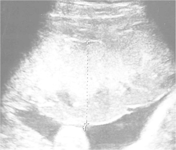
Purpose: The aim of this study is to evaluate the efficacy of an ultrasonographic measurement of placental thickness and the correlation of a thick placenta with adverse perinatal outcome.
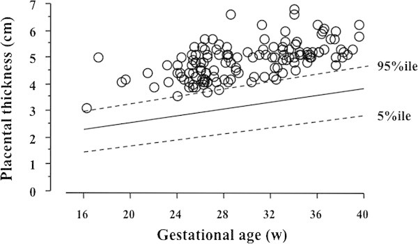
Methods: Placental thickness was measured in single gravidas, 16 to 40 weeks of gestation, between 2005 and 2009. Placentas were considered to be thick if their measured thickness were above the 95th percentile for gestational age.
Results: The incidence of thick placentas was 4.3% (138/3,183). Perinatal morbidity and neonatal conditions were worse in cases with thick placenta rather than without thick placenta.
Conclusions: Ultrasonographic measurement of placental thickness is a simple method to estimate placental size. Thick placenta may be a useful predictor of adverse pregnancy outcomes.
Keywords: Measurement of placental thickness; Perinatal outcome; Thick placenta; Ultrasonography.
Figures
References
-
- Bleker OP, Kloosterman GJ, Breur W, Mieras DJ. The volumetric growth of the human placenta: a longitudinal ultrasonic study. Am J Obstet Gynecol. 1977;127:657–661. - PubMed
-
- de Paula CF, Ruano R, Campos JA, Zugaib M. Placental volumes measured by 3-dimensional ultrasonography in normal pregnancies from 12 to 40 weeks’ gestation. J Ultrasound Med. 2008;27:1583–1590. - PubMed
LinkOut - more resources
Full Text Sources
Other Literature Sources
