. 2014 Jul 31;9(7):e103655.
doi: 10.1371/journal.pone.0103655. eCollection 2014.

Polarized axonal surface expression of neuronal KCNQ potassium channels is regulated by calmodulin interaction with KCNQ2 subunit

Affiliations

Polarized axonal surface expression of neuronal KCNQ potassium channels is regulated by calmodulin interaction with KCNQ2 subunit

John P Cavaretta et al. PLoS One. .

Abstract

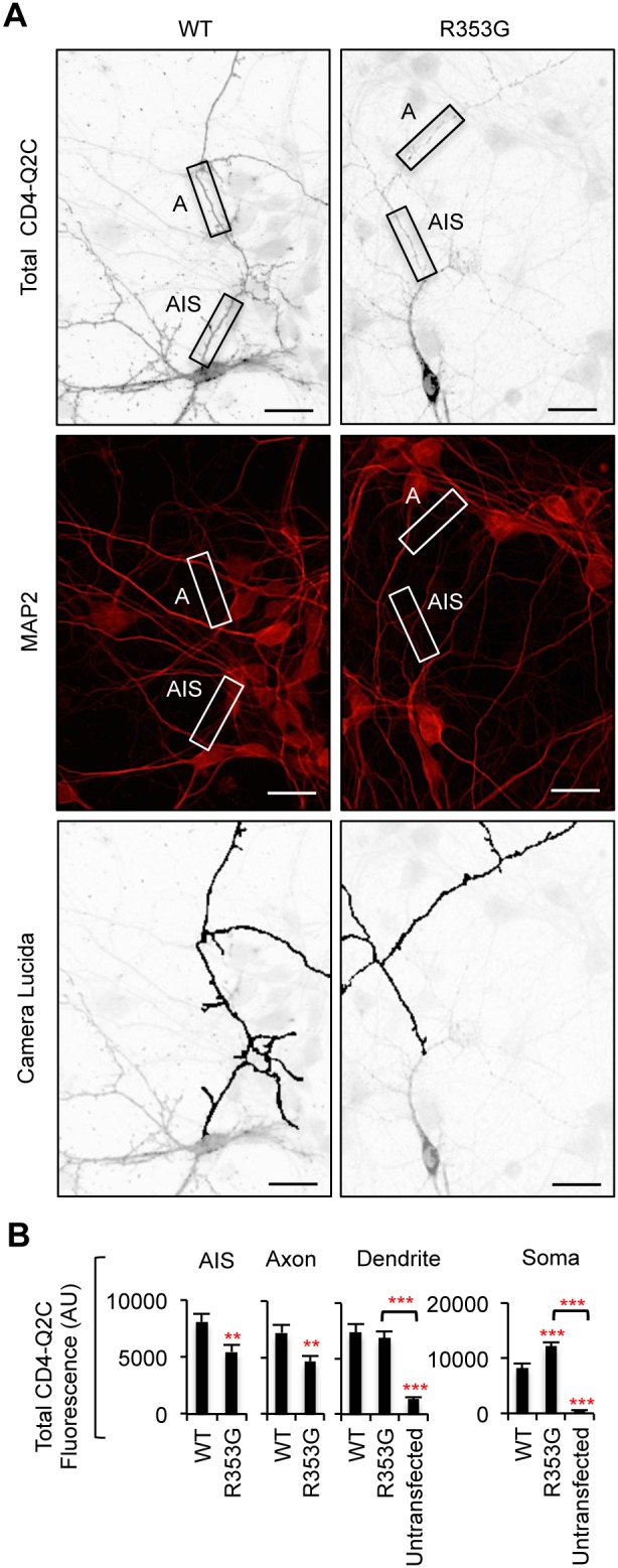
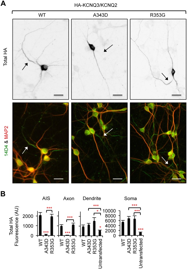
KCNQ potassium channels composed of KCNQ2 and KCNQ3 subunits give rise to the M-current, a slow-activating and non-inactivating voltage-dependent potassium current that limits repetitive firing of action potentials. KCNQ channels are enriched at the surface of axons and axonal initial segments, the sites for action potential generation and modulation. Their enrichment at the axonal surface is impaired by mutations in KCNQ2 carboxy-terminal tail that cause benign familial neonatal convulsion and myokymia, suggesting that their correct surface distribution and density at the axon is crucial for control of neuronal excitability. However, the molecular mechanisms responsible for regulating enrichment of KCNQ channels at the neuronal axon remain elusive. Here, we show that enrichment of KCNQ channels at the axonal surface of dissociated rat hippocampal cultured neurons is regulated by ubiquitous calcium sensor calmodulin. Using immunocytochemistry and the cluster of differentiation 4 (CD4) membrane protein as a trafficking reporter, we demonstrate that fusion of KCNQ2 carboxy-terminal tail is sufficient to target CD4 protein to the axonal surface whereas inhibition of calmodulin binding to KCNQ2 abolishes axonal surface expression of CD4 fusion proteins by retaining them in the endoplasmic reticulum. Disruption of calmodulin binding to KCNQ2 also impairs enrichment of heteromeric KCNQ2/KCNQ3 channels at the axonal surface by blocking their trafficking from the endoplasmic reticulum to the axon. Consistently, hippocampal neuronal excitability is dampened by transient expression of wild-type KCNQ2 but not mutant KCNQ2 deficient in calmodulin binding. Furthermore, coexpression of mutant calmodulin, which can interact with KCNQ2/KCNQ3 channels but not calcium, reduces but does not abolish their enrichment at the axonal surface, suggesting that apo calmodulin but not calcium-bound calmodulin is necessary for their preferential targeting to the axonal surface. These findings collectively reveal calmodulin as a critical player that modulates trafficking and enrichment of KCNQ channels at the neuronal axon.


Conflict of interest statement

Competing Interests: The authors have declared that no competing interests exist.

Figures

Figure 1
Figure 1. Enrichment of HA-KCNQ3/KCNQ2 channels and CD4-Q2C at the axonal surface.
Schematic drawings (not to scale) of a human KCNQ2 subunit (accession #Y15065), including the subunit interaction domain (Sid, amino acids 580–623) , , and CaM-binding domain (amino acids 323–579) , . (B) Immunoblot analysis of CaM in cultured rat hippocampal neurons at 5–9 days in vitro (DIV). Glyceraldehyde-3-phosphate dehydrogenase (GAPDH) served as a loading control. (C) Schematic drawings (not to scale) of a human CD4 protein, and CD4 fused to KCNQ2 C-terminal tail (CD4-Q2C, [18]). (D) Surface immunostaining of hippocampal neurons (DIV 7) transfected with KCNQ2 and KCNQ3 containing an extracellular hemagglutinin (HA) epitope (HA-KCNQ3). Neuronal soma and dendrites were visualized by immunostaining for MAP2. Surface HA-KCNQ3/KCNQ2 channels are enriched on a MAP2-negative neurite that originates directly from the soma. (E) Pseudo-color image of the inset in Fig. 1D displays differences in the surface HA intensity. Surface HA-KCNQ3/KCNQ2 channels are enriched at the initial segment of an axon. (F) Surface immunostaining of hippocampal neurons (DIV 8) transfected with CD4, or CD4-Q2C. (G) Pseudo-color images of the insets in Fig. 1F display differences in the surface CD4 intensity. Fusion of KCNQ2 C-terminal tail enriches CD4 at the axonal surface. Camera lucida drawings of the neuronal images (D, F) show the soma and dendrites (gray) and an axon (black). Arrows mark the main axon. Scale bars are 20 µm.
Figure 2
Figure 2. Mutations in IQ motif abolish axonal enrichment of surface CD4-Q2C.
(A) Schematic drawings (not to scale) of a human KCNQ2 subunit (accession #Y15065) and CD4-Q2C showing CaM-binding domain. The amino acids in bold text are critical residues in the CaM-binding consensus IQ motif in helix A. Mutations in the underlined amino acids have been shown to abolish (L339R, I340E, and A343D) or moderately decrease (R353G) CaM interaction with KCNQ2 , . Mutations in the amino acids colored red are associated with BFNC . (B) Lysates from HEK293T cells expressing CaM and CD4-Q2C wild-type (WT) or mutant proteins (L339R, I340E, and A343D) were subjected to immunoprecipitation (IP) with the CD4 antibody. Immunoprecipitation and total cell lysates were analyzed by immunoblotting for CD4 and CaM. β-actin served as a loading control. The L339R, I340E, and A343D mutations abolished co-immunoprecipitation of CaM with CD4-Q2C. (C) Surface immunostaining of CD4-Q2C WT or mutant proteins in hippocampal neurons (DIV 7–8). Camera lucida drawings (lower) of the inverted images of surface CD4-Q2C (upper) show the soma and dendrites (gray) and an axon (black). The axon was identified by immunostaining for the AIS marker phospho IκBα Ser32 (14D4) in the neurons cotransfected with GFP, which allows visualization of all neurites (Figure S3). Arrows indicate the AIS. Scale bars are 20 µm. The L339R, I340E, and A343D mutations abolished surface expression of CD4-Q2C at the AIS and distal axon. (D) The surface “Axon/Dendrite” ratios of CD4-Q2C were reduced to nearly 0 by L339R, I340E, and A343D mutations. (E) Background subtracted, mean intensity of surface CD4 fluorescence in the AIS, distal axons, soma, and major dendrites. AU, arbitrary unit. The sample number for each construct used in (D, E) was as follows: WT (n = 18), L339R (n = 20), I340E (n = 12), A343D (n = 25), and untransfected (n = 20). Ave ± SEM (*p<0.05, **p<0.01, ***p<0.001).
Figure 3
Figure 3. Mutations in IQ motif block CD4-Q2C expression in the axon.
(A) Permeabilized immunostaining of CD4-Q2C wild-type (WT) or mutant (L339R, I340E, or A343D) in GFP-cotransfected hippocampal neurons (DIV 7–8). Camera lucida drawings (lower) of the inverted images of total CD4-Q2C (upper) show the soma and dendrites (gray) and an axon (black). Arrows indicate the AIS identified by phospho IκBα Ser32 (14D4) immunostaining. The L339R, I340E, and A343D mutations abolished total (surface and intracellular) expression of CD4-Q2C in the axon. (B) Background subtracted, mean intensity of total CD4 fluorescence in the AIS, distal axons, soma, and major dendrites. AU, arbitrary unit. The sample number for each construct used was as follows: WT (n = 18), L339R (n = 20), I340E (n = 12), A343D (n = 25), and untransfected (n = 20). Ave ± SEM (*p<0.05, **p<0.01, ***p<0.001). (C) Surface expression of A343D mutant CD4Q2C was absent (upper panel) although total (surface and intracellular) expression of A343D mutant CD4Q2C was evident in the MAP2-positive soma and proximal dendrites (lower panel). (A, C) Scale bars are 20 µm.
Figure 4
Figure 4. The BFNC R353G mutation blocks axonal enrichment of surface CD4-Q2C.
(A) The R353G mutation reduced but did not abolish co-immunoprecipitation of CaM with CD4-Q2C from transfected HEK293T cells. β-actin served as a loading control for total cell lysates (B) Surface immunostaining of WT or R353G mutant CD4-Q2C in hippocampal neurons (DIV 7–8). Camera lucida drawings (lower) of the inverted images of surface CD4-Q2C (upper) show the soma and dendrites (gray) and an axon (black). The axon was identified by the lack of MAP2 immunostaining in the neurons cotransfected with GFP. The R353G mutation blocked enrichment of CD4-Q2C on the axonal surface by increasing its somatodendritic surface expression. Arrows mark the main axon. Scale bars are 20 µm. (C) In comparison to WT, the surface “Axon/Dendrite” fluorescence ratio of CD4-Q2C was reduced to 1 by the R353G mutation, whereas the surface “AIS/Axon” ratio was unaffected. (D) Background subtracted, mean intensity of surface CD4 fluorescence in the AIS, distal axons, soma, and major dendrites. The R353G mutation increased CD4-Q2C expression at the somatodendritic surface compared to WT. The sample number for each construct used in (C, D) was as follows: WT (n = 23), R353G (n = 18), and untransfected (n = 15). AU, arbitrary unit. Ave ± SEM (*p<0.05, **p<0.01, ***p<0.001).
Figure 5
Figure 5. The R353G mutation reduces CD4-Q2C expression in the axon.
(A) Permeabilized immunostaining of wild-type (WT) or R353G mutant CD4-Q2C in hippocampal neurons (DIV 7–8) cotransfected with GFP. Neuronal soma and dendrites were identified by MAP2 immunostaining (middle). The insets in the representative inverted images of total CD4-Q2C show the initial segment (AIS) and distal segment (A) of the MAP2-negative axon in transfected neurons. Camera lucida drawings (lower) of the neuronal images (upper) show the soma and dendrites (gray) and an axon (black). The R353G mutation reduced but did not abolish total (surface and intracellular) expression of CD4-Q2C from the axon. Scale bars are 20 µm. (B) Background subtracted, mean intensity of total CD4 fluorescence in the AIS, distal axons, soma, and major dendrites. AU, arbitrary unit. The sample number for each construct used was as follows: WT (n = 23), R353G (n = 18), and untransfected (n = 15). AU, arbitrary unit. Ave ± SEM (**p<0.01, ***p<0.001).
Figure 6
Figure 6. CD4-Q2C accumulates in the ER after BFA treatment.
(A) Schematic drawings of a hippocampal neuron containing a continuous network of the ER from the soma to the dendrites. Treatment with brefeldin-A (BFA) leads to inhibition of anterograde transport from the ER to the Golgi complex . (B–C) The cultured hippocampal neurons (DIV 5) were treated with vehicle control (No BFA) or BFA (0.75 µg/ml) at 30 min post transfection with GFP and CD4-Q2C wild-type (WT) (B), I340E mutant (C), or R353G mutant (D). At 16 hr post-BFA treatment, permeabilized immunostaining was performed to visualize total (surface and intracellular) expression of CD4-Q2C (inverted images, upper). BFA treatment caused newly synthesized wild-type and all mutant proteins to accumulate at perinuclear regions in the soma and proximal dendrites. Arrows mark the main axon identified by the lack of MAP2 immunostaining in the neurons cotransfected with GFP. Camera lucida drawings (lower) show the soma and dendrites (gray) and an axon (black). Scale bars are 20 µm.
Figure 7
Figure 7. The I340E mutation impairs CD4-Q2C trafficking from the ER to the axon.
Pulse-chase assay of wild-type (WT) or mutant (I340E and R353G) CD4-Q2C proteins after BFA washout. At 16 hr post BFA treatment, BFA was washed out and the neurons were placed in the cell culture incubator for 0, 1, 2, 4, and 8 hr. Permeabilized immunostaining was performed to visualize total (surface and intracellular) expression of CD4-Q2C in neurons cotransfected with GFP. The axon was identified by immunostaining with the AIS marker phospho IκBα Ser32 (14D4). (A) Representative images of CD4-Q2C WT or mutant proteins at 0, 4, and 8 hr after BFA washout. Camera lucida drawings (middle) of the GFP-transfected neurons (upper) show the soma and dendrites (gray) and an axon (black). Scale bars in the upper and middle panels are 10 µm. The small lower panels are representative inverted images of CD4-Q2C in the AIS (AIS), distal axons (A), and dendrites (D) in transfected neurons (insets). Scale bars of the small lower panels are 5 µm. (B) Schematic drawings of a hippocampal neuron containing CD4-Q2C after BFA washout. (C) Background subtracted, mean intensity of total CD4 fluorescence in the AIS, distal axons, and major dendrites. The wild-type CD4-Q2C proteins gradually appeared at the AIS and distal axons upon BFA washout. The I340E mutation abolished whereas the R353G mutation reduced the accumulation of CD4-Q2C at the AIS and distal axons. The sample numbers per time point for each construct (n = 8–21). AU, arbitrary unit. Ave ± SEM (*p<0.05, **p<0.01, ***p<0.001).
Figure 8
Figure 8. The A343D mutation blocks axonal enrichment of surface HA-KCNQ3/KCNQ2 channels.
(A) Lysates from HEK293T cells expressing CaM and wild-type KCNQ2 (WT) or mutant KCNQ2 (A343D and R353G) were subjected to immunoprecipitation (IP) with the KCNQ2 antibody. Immunoprecipitation and total cell lysates were analyzed by immunoblotting for KCNQ2 and CaM. β-actin served as a loading control. The A343D mutation abolished whereas the R353G modestly reduced co-immunoprecipitation of CaM with KCNQ2. (B) Representative inverted images of surface HA-KCNQ3 in hippocampal neurons cotransfected with GFP and KCNQ2 WT or mutants (A343D and R353G). The A343D but not the R353G mutation abolished surface expression of HA-KCNQ3/KCNQ2 at the axon, which was identified by immunostaining for the AIS marker, phospho IκBα Ser32 (14D4) (Figure S4). Camera lucida drawings (middle) of neuronal images (upper) show the soma and dendrites (gray) and an axon (black). Pseudo-color images (lower) of the insets in the neuronal images (upper) display differences in the surface HA intensity. Arrows indicate the AIS. Arrowheads mark another axon. Scale bars: 20 µm. (C) The surface “Axon/Dendrite” ratio was reduced by the A343D but not the R353G mutation. The surface AIS/distal axon ratio for A343D mutant channels was not calculated due to their absence at the axonal and AIS surface. (D) Background subtracted, mean intensity of surface HA fluorescence in the AIS, distal axons, soma, and major dendrites. The sample number for each construct was as follows: WT (n = 27), A343D (n = 22), R353G (n = 21), and untransfected (n = 15). AU, arbitrary unit. Ave ± SEM (*p<0.05, **p<0.01, ***p<0.001).
Figure 9
Figure 9. The A343D mutation abolishes axonal expression of HA-KCNQ3/KCNQ2 channels.
(A) Permeabilized immunostaining was performed to visualize total (surface and intracellular) expression of HA-KCNQ3/KCNQ2 WT or mutant (A343D and R353G) channels (inverted images, upper). The axon was identified by the AIS marker phospho IκBα Ser32 (14D4) whereas neuronal soma and dendrites were visualized by MAP2 immunostaining (lower). The A343D but not the R353G mutation abolished total expression of HA-KCNQ3/KCNQ2 channels at the axon. Arrows indicate the AIS. Scale bars are 20 µm. (B) Background subtracted, mean intensity of total HA fluorescence in the AIS, distal axons, soma, and major dendrites. The sample number for each construct was as follows: WT (n = 27), A343D (n = 22), R353G (n = 21), and untransfected (n = 15). AU, arbitrary unit. Ave ± SEM (*p<0.05, ***p<0.001).
Figure 10
Figure 10. The HA-KCNQ3/KCNQ2-A343D mutant channels are absent from the ER-negative axon.
(A) Permeabilized immunostaining of HA-KCNQ3/KCNQ2 channels (inverted images, left) and MAP2 (middle) were performed in cultured hippocampal neurons (DIV 7) cotransfected with the CD4 proteins harboring the ER retention/retrieval motif (CD4-KDEL, green). The wild type (WT) and R353G mutant channels were found at the axons where the A343D mutant channels and CD4-KDEL proteins were absent. The insets show the major axon. (B) The cultured hippocampal neurons (DIV 5) were treated with vehicle control (No BFA) or BFA (0.75 µg/ml) at 30 min post transfection with HA-KCNQ3 and KCNQ2 wild-type (WT), or mutant (A343D and R353G). At 16 hr post-BFA treatment, permeabilized immunostaining was performed to visualize total (surface and intracellular) expression of HA-KCNQ3/KCNQ2 channels. The axon was identified by the AIS marker phospho IκBα Ser32 (14D4), whereas neuronal soma and dendrites were visualized by MAP2 immunostaining. BFA treatment caused newly synthesized wild-type and all mutant channels to accumulate at perinuclear regions in the soma and dendrites but not axons. Arrows mark the AIS. Scale bars are 20 µm.
Figure 11
Figure 11. The A343D mutation impairs HA-KCNQ3/KCNQ2 trafficking from the ER to the axon.
(A–B) Pulse-chase assay of wild-type (WT) or mutant (A343D and R353G) HA-KCNQ3/KCNQ2 channels after BFA washout. Permeabilized immunostaining was performed to visualize total (surface and intracellular) expression of HA-KCNQ3/KCNQ2 channels in the soma and AIS (A) as well as in the distal axons and dendrites (B) at indicated time points post-BFA removal (inverted images). The axon was identified by the AIS marker phospho IκBα Ser32 (14D4), whereas neuronal soma and dendrites were visualized by MAP2 immunostaining. Arrows indicate the AIS. Scale bars: 10 µm. (C) Background subtracted, mean intensity of total HA fluorescence in the AIS, distal axon, and major dendrites. Upon BFA removal, the A343D mutation but not the R353G mutation markedly reduced the appearance of HA-KCNQ3/KCNQ2 at the AIS and distal axon for the duration of the 8 hr BFA washout. The sample numbers were (n = 8–22) per time point for each construct. AU, arbitrary unit. Ave ± SEM (*p<0.05, **p<0.01, ***p<0.001).
Figure 12
Figure 12. Expression of wild-type KCNQ2 but not KCNQ2-A343D or KCNQ2-R353G decreases neuronal excitability.
(A) Hypothesis by which exogeneously expressed KCNQ2 subunits affect neuronal excitability. At an early stage of hippocampal culture, the level of endogenous KCNQ2/KCNQ3 channels is low . Transfection of wild-type KCNQ2 or R353G mutant KCNQ2 would reduce action potential firing by increasing axonal surface expression of KCNQ2-containing homomeric channels and heteromeric channels formed with endogenous KCNQ3 subunits. In contrast, expression of A343D mutant KCNQ2 subunits deficient in CaM binding would have little effect on action potential firing due to their inability to exit the ER and express on the axonal surface. (B–D) Whole-cell patch clamp recording in cultured hippocampal neurons (DIV 6–8) that were cotransfected with GFP and KCNQ2 WT or mutants (A343D and R353G). Spike trains were evoked in untransfected or GFP-positive pyramidal neurons by delivering constant somatic current pulses for 500 ms duration at a resting potential of –60 mV. (B) Representative traces of action potentials induced by 100 pA injection. (C) Average instantaneous AP firing rates (Hz) induced by 0–200 pA injection into untransfected neurons (n = 10), or neurons transfected with GFP (n = 10), GFP + KCNQ2 WT (n = 10), GFP + KCNQ2 A343D (n = 10), or GFP + KCNQ2 R353G (n = 9). (D) Summary plots illustrating the effect of transfection on instantaneous firing rates at 100 pA. Ave ± SEM (*p<0.05 for untransfected vs. WT, #p<0.05 for GFP vs. WT, ∧p<0.05 for WT vs. A343D).
Figure 13
Figure 13. CaM1234 reduces enrichment of HA-KCNQ3/KCNQ2 channels at the axonal surface.
(A) Surface expression of HA-KCNQ3/KCNQ2 channels in hippocampal neurons (DIV 7–8) cotransfected with empty vector (pcDNA3), CaM wild-type (WT) or Ca2+-insensitive mutant CaM (CaM1234). Endogenous and transfected CaM proteins were immunostained with anti-CaM antibodies (CaM, lower inverted images). HA-KCNQ3/KCNQ2 channels were enriched at the axonal surface in the presence of CaM WT or CaM1234. Arrows mark the main axon. Scale bars are 20 µm. (B) Overexpression of CaM WT or CaM1234 did not grossly affect neuronal polarity as indicated by immunostaining of the somatodendritic marker MAP2 and the AIS marker ankryin-G. Arrows indicate the AIS. Scale bars are 20 µm. (C) The surface “Axon/Dendrite” ratio was reduced by 33% by coexpression with CaM1234 (n = 21) compared to coexpression with CaM WT (n = 33) or empty vector pcDNA3 (n = 43). The surface “AIS/Axon” ratio of CD4-Q2C was increased by coexpression with CaM1234 compared to CaM WT but not pcDNA3. (D) Background subtracted, mean intensity of the CaM fluorescence in the soma of transfected and untransfected neurons (n = 23). (E) Background subtracted, mean intensity of surface and total (surface and intracellular) HA fluorescence in transfected and untransfected neurons. CaM1234 modestly decreased surface expression of HA-KCNQ3/KCNQ2 channels at the AIS and axon. (D, E) AU, arbitrary unit. Ave ± SEM (*p<0.05, **p<0.01, ***p<0.001). (F) Model by which apoCaM interaction with helices A and B of KCNQ2 and KCNQ3 is critical for trafficking of KCNQ2/KCNQ3 channels from the ER to the axonal surface.

References

    1. Brown DA, Passmore GM (2009) Neural KCNQ (Kv7) channels. Br J Pharmacol 156: 1185–1195. - PMC - PubMed
    1. Wang HS, Pan Z, Shi W, Brown BS, Wymore RS, et al. (1998) KCNQ2 and KCNQ3 potassium channel subunits: molecular correlates of the M-channel. Science 282: 1890–1893. - PubMed
    1. Cooper EC, Harrington E, Jan YN, Jan LY (2001) M channel KCNQ2 subunits are localized to key sites for control of neuronal network oscillations and synchronization in mouse brain. J Neurosci 21: 9529–9540. - PMC - PubMed
    1. Roche JP, Westenbroek R, Sorom AJ, Hille B, Mackie K, et al. (2002) Antibodies and a cysteine-modifying reagent show correspondence of M current in neurons to KCNQ2 and KCNQ3 K+ channels. Br J Pharmacol 137: 1173–1186. - PMC - PubMed
    1. Devaux JJ, Kleopa KA, Cooper EC, Scherer SS (2004) KCNQ2 is a nodal K+ channel. J Neurosci 24: 1236–1244. - PMC - PubMed

Publication types