

Anti-hepatotoxic activities of Hibiscus sabdariffa L. in animal model of streptozotocin diabetes-induced liver damage
- PMID: 25077880
- PMCID: PMC4131030
- DOI: 10.1186/1472-6882-14-277
Anti-hepatotoxic activities of Hibiscus sabdariffa L. in animal model of streptozotocin diabetes-induced liver damage
Abstract
Background: Flavonoid-rich aqueous fraction of methanolic extract of Hibiscus sabdariffa calyx was evaluated for its anti-hepatotoxic activities in streptozotocin-induced diabetic Wistar rats.
Methods: Diabetes Mellitus was induced in Wistar rats by a single i.p injection of 80 mg/kg b.w. streptozotocin (STZ) dissolved in 0.1 M citrate buffer (pH 6.3).
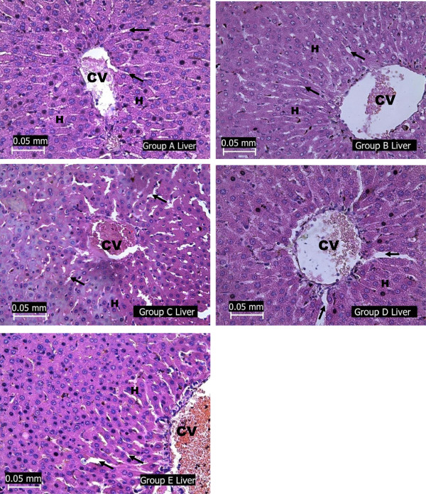
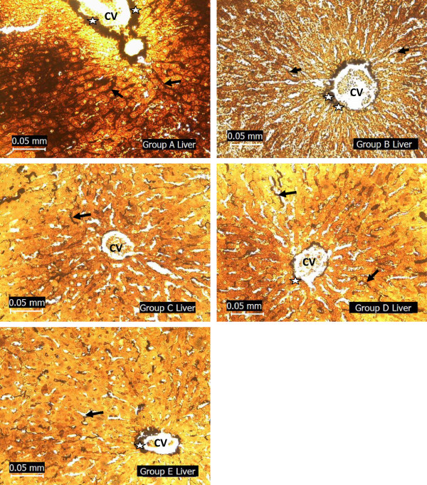
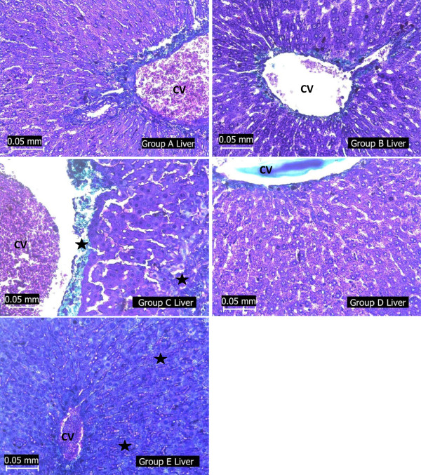
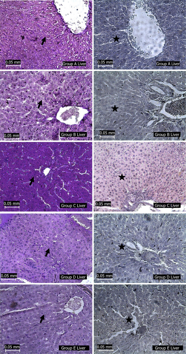
Results: The ameliorative effects of the extract on STZ-diabetes induced liver damage was evident from the histopathological analysis and the biochemical parameters evaluated in the serum and liver homogenates. Reduced levels of glutathione (GSH), catalase (CAT), superoxide dismutase (SOD), and glutathione peroxidase (GPx) (3.76 ± 0.38 μM, 0.42 ± 0.04 U/L, 41.08 ± 3.04 U/ml, 0.82 ± 0.04 U/L respectively) in the liver of diabetic rats were restored to a near normal level in the Hibiscus sabdariffa-treated rats (6.87 ± 0.51 μM, 0.72 ± 0.06 U/L, 87.92 ± 5.26 U/ml, 1.37 ± 0.06 U/L respectively). Elevated levels of aspartate amino transferase (AST), alanine amino transferase (ALT) and alkaline phosphatase (ALP) in the serum of diabetic rats were also restored in Hibiscus sabdariffa -treated rats. Examination of stained liver sections revealed hepatic fibrosis and excessive glycogen deposition in the diabetic rats. These pathological changes were ameliorated in the extract-treated rats.
Conclusion: The anti-hepatotoxic activity of Hibiscus sabdariffa extract in STZ diabetic rats could be partly related to its antioxidant activity and the presence of flavonnoids.
Figures
References
-
- Levinthal GN, Tavill AS. Liver disease and diabetes mellitus. Clin Diabetes. 1999;17(2):73–93.
-
- Subramoniam A, Pushpangadan P. Development of phytomedicine for liver diseases. Ind J Pharmacol. 1999;31:166–175.
-
- Muhtaseb MS, El Talwar D, Duncan A, St J, O'reilly D, Mckee RF, Anderson JH, Foulisa FIG. Free radical activity and lipid soluble anti-oxidant vitamin status in patients with long-term ileal pouch-anal anastomosis. Colorectal Dis. 2008;11:67–72. - PubMed
-
- Ishibashi H, Nakamura M, Komori A, Migita K, Shimoda S. Liver architecture, cell function, and disease. Semin Immunopathol. 2009;31:399–409. - PubMed
Pre-publication history
-
- The pre-publication history for this paper can be accessed here:http://www.biomedcentral.com/1472-6882/14/277/prepub
MeSH terms
Substances
LinkOut - more resources
Full Text Sources
Other Literature Sources
Medical
Miscellaneous
