. 2014 Oct 3;289(40):27551-61.
doi: 10.1074/jbc.M113.528174. Epub 2014 Jul 30.

Endoplasmic reticulum stress is chronically activated in chronic pancreatitis

Affiliations

Endoplasmic reticulum stress is chronically activated in chronic pancreatitis

Raghuwansh P Sah et al. J Biol Chem. .

Abstract

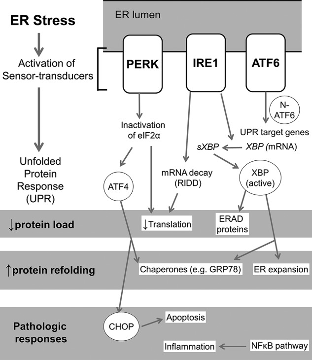
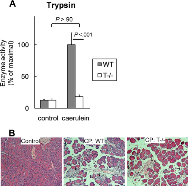
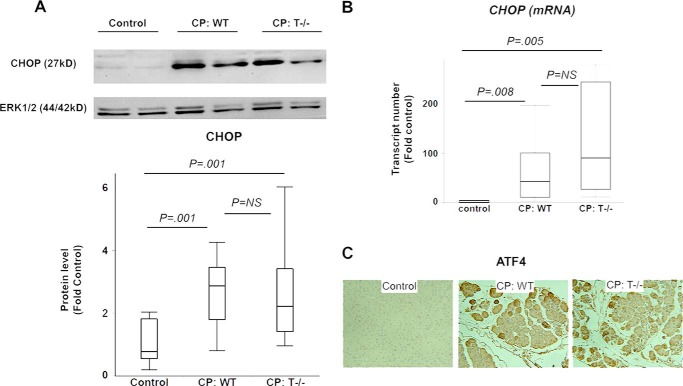
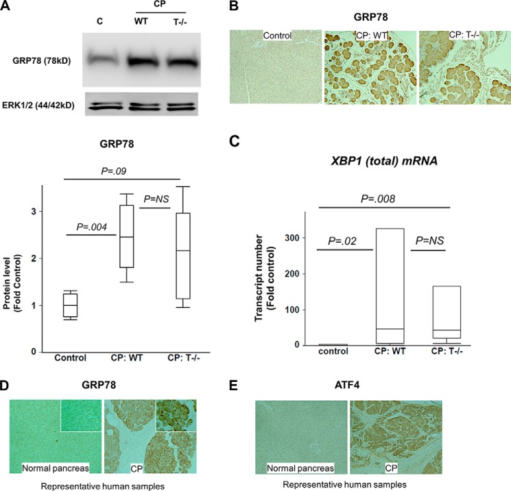
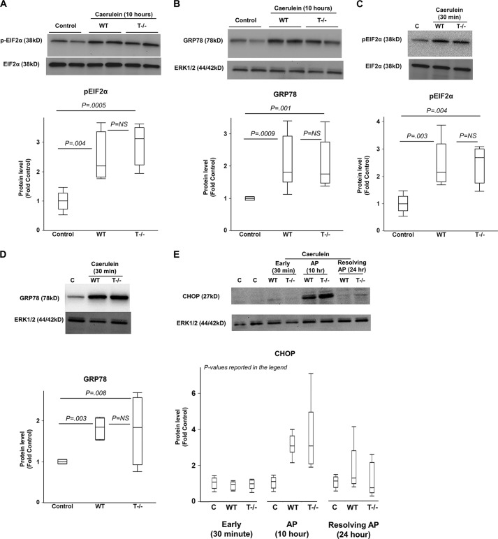
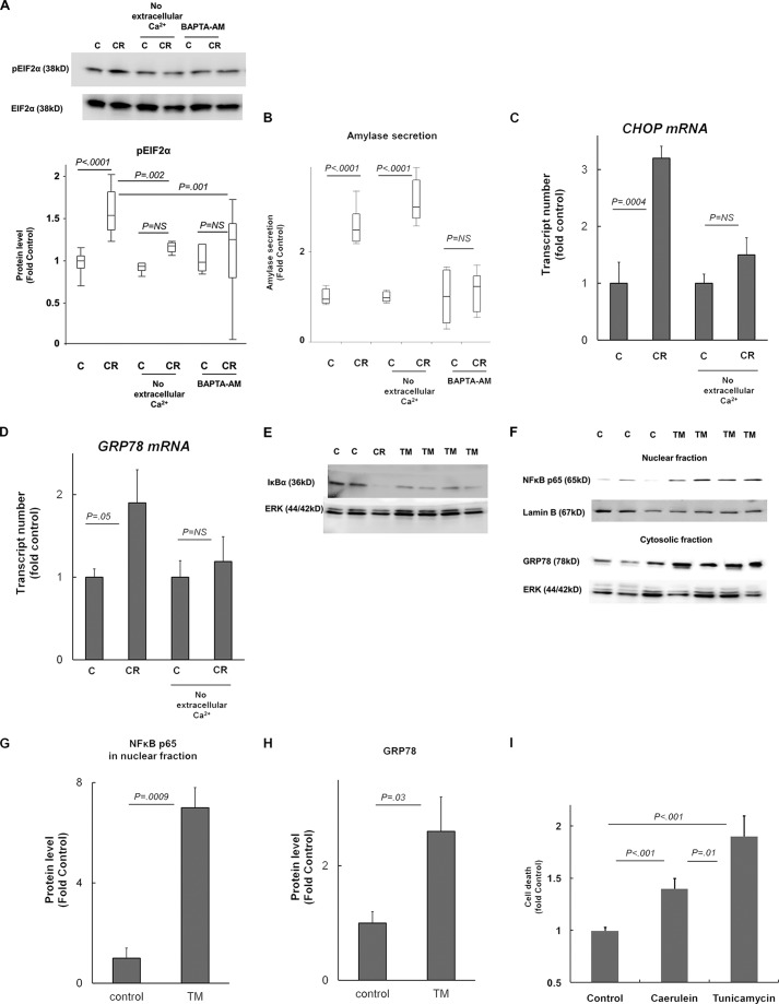
The pathogenesis of chronic pancreatitis (CP) is poorly understood. Endoplasmic reticulum (ER) stress has now been recognized as a pathogenic event in many chronic diseases. However, ER stress has not been studied in CP, although pancreatic acinar cells seem to be especially vulnerable to ER dysfunction because of their dependence on high ER volume and functionality. Here, we aim to investigate ER stress in CP, study its pathogenesis in relation to trypsinogen activation (widely regarded as the key event of pancreatitis), and explore its mechanism, time course, and downstream consequences during pancreatic injury. CP was induced in mice by repeated episodes of acute pancreatitis (AP) based on caerulein hyperstimulation. ER stress leads to activation of unfolded protein response components that were measured in CP and AP. We show sustained up-regulation of unfolded protein response components ATF4, CHOP, GRP78, and XBP1 in CP. Overexpression of GRP78 and ATF4 in human CP confirmed the experimental findings. We used novel trypsinogen-7 knock-out mice (T(-/-)), which lack intra-acinar trypsinogen activation, to clarify the relationship of ER stress to intra-acinar trypsinogen activation in pancreatic injury. Comparable activation of ER stress was seen in wild type and T(-/-) mice. Induction of ER stress occurred through pathologic calcium signaling very early in the course of pancreatic injury. Our results establish that ER stress is chronically activated in CP and is induced early in pancreatic injury through pathologic calcium signaling independent of trypsinogen activation. ER stress may be an important pathogenic mechanism in pancreatitis that needs to be explored in future studies.

Keywords: Chronic Pancreatitis; ER Stress; Endoplasmic Reticulum (ER); NF-kappa B (NF-KB); Pancreas; Trypsinogen Activation; Unfolded Protein Response (UPR).


Figures

FIGURE 1.
FIGURE 1.
ER stress and UPR pathways. ER stress sensing triggers a) auto-phosphorylation of PERK; b) oligomerization, auto-phosphorylation, and activation of ribonuclease activity of IRE-1; and c) migration of ATF6 to Golgi resulting in its cleavage and liberation of the N-terminal cytosolic fragment, which translocates to the nucleus (8). Reduction in translation occurs due to inactivation of EIF2α after undergoing phosphorylation by pPERK, as well as due to degradation of any mRNA in the vicinity of activated IRE-1 by its nonspecific ribonuclease activity (termed regulated IRE1-dependent degradation (RIDD)) (8). IRE-1 has a specific ribonuclease activity resulting in targeted splicing of XBP-1 transcript (sXBP1), which is translated into active transcription factor XBP-1 (8, 29). XBP-1 leads to synthesis of a) chaperones such as GRP78; b) refolding proteins such as protein disulfide isomerase; c) endoplasmic reticulum-associated degradation (ERAD) products; and d) lipids for the expansion of ER membrane (8). Transcription of XBP-1 is induced by nuclear N-terminal fragment of ATF6 (29). Selective translation of ATF-4 occurs in low EIF2α abundance, which induces transcription of CHOP and GADD34. The initial UPR responses are aimed at restoring ER homeostasis. Pathogenic responses result when homeostasis fails due to unabated ER stress. Significant redundancies and overlaps occur in each of the three pathways; their relative roles may be specific to cell type and context. ER stress- and UPR-related downstream transcription factors are shown in circles.
FIGURE 2.
FIGURE 2.
A, T−/− mice lack significant intra-acinar trypsinogen activation. Trypsin activity was measured in pancreatic homogenates 30 min after caerulein injection and reported as the percentage of maximal activity relative to the respective controls (saline injected). n = 10–14/group. B, CP was comparable in WT and T−/− mice. Representative hematoxylin and eosin-stained sections (100×) from control and CP groups demonstrating similar histomorphologic features of acinar atrophy (white arrows), ductular metaplasia (*), and chronic inflammatory infiltrate (black arrows) in WT and T−/− mice (n = 20/group in two independent experiments) are shown. A significant proportion of the empty spaces in the sections is composed of fibrotic areas that can be directly visualized with Sirius red stain as reported elsewhere (6). Error bars indicate means ± S.E.
FIGURE 3.
FIGURE 3.
A and B, overexpression of CHOP in CP. A, CHOP protein was up-regulated in WT and T−/− mice with CP. B, CHOP mRNA up-regulation in CP. Wilcoxon test used for analysis. n = 6/group. C, overexpression of ATF4 in CP. Representative ATF4 immunostained sections (100×) demonstrating higher ATF4 positivity in WT and T−/− mice with CP. n = 4–5/group. Error bars indicate means ± S.E. NS, not significant.
FIGURE 4.
FIGURE 4.
A and B, overexpression of GRP78 in CP. A, Western blot showing GRP78 up-regulation in WT and T−/− mice with CP. n = 5/group. C lane, control. B, immunostaining for GRP78 demonstrates up-regulation in CP, comparable in WT and T−/− mice. Representative sections are shown (n = 4–5/group). C, up-regulation of XBP1(total) mRNA in CP. Wilcoxon test used for analysis. n = 6/group. D and E, sustained ER stress in human CP. GRP78 (D) and ATF4 (E) immunostaining demonstrates up-regulation in human CP as compared with normal human pancreas confirming sustained ER stress in human CP. Representative sections (100×) are shown. Inset in D shows zoomed in (300×) views. Error bars indicate means ± S.E. NS, not significant.
FIGURE 5.
FIGURE 5.
A and B, ER stress and UPR activation occurs in AP independent of trypsinogen activation. Proteins were analyzed by Western blot in pancreatic homogenates from mice with AP after hourly caerulein injections 10 times as compared with saline-injected controls. A, phosphorylation of eIF2α during AP in WT and T−/− mice. n = 4/group. B, up-regulation of GRP78 during AP in WT and T−/− mice. n = 7–10/group. C and D, ER stress induction occurred very early in the course of pancreatic injury independent of trypsinogen activation. Proteins analyzed by Western blot in pancreatic homogenates from mice 30 min after caerulein injection as compared with saline-injected controls. Phosphorylation of eIF2α is shown in C. C lane, control. Up-regulation of GRP78 is shown in D. n = 4–5/group. E, time course of CHOP activation during acute pancreatic injury. Early time point (30 min after caerulein injection), AP time point (after hourly caerulein injection 10 times), and time point of resolution of AP (mice sacrificed after 16 h of observation following hourly caerulein injection eight times) have been shown. CHOP activation was seen during the AP time point but was absent during the resolution phase as well as at the early stage of pancreatic injury. p values for WT versus control, T−/− versus control, and WT versus T−/−, respectively: < 0.0001, < 0.0001, and not significant at AP time point (n = 9–10/group); each not significant at 30 min time point (n = 5/group) and resolving AP time point (n = 5/group). Error bars indicate means ± S.E. NS, not significant.
FIGURE 6.
FIGURE 6.
A–D, ER stress is mediated by pathologic Ca2+ signaling during early pancreatic injury. Mice acini were incubated with 100 nm caerulein (CR) or control (C) for 30 min (A and B) or 2 h (C and D) in HEPES containing 1 mm Ca2+ or without Ca2+ (no extracellular Ca2+ group). A and B, in the BAPTA-AM group, acini were preincubated with 10 μm BAPTA-AM in HEPES buffer lacking Ca2+ for 30 min prior to treatment with caerulein in HEPES containing 1 mm Ca2+. All experiments were conducted in parallel, and data shown represent n = 6–8/group pooled from two independent experiments. Amylase secretion was measured in supernatants after completion of incubation in the same experiments described above and was normalized to total protein. Amylase secretion data are intended to verify the internal validity of the experimental conditions. C and D, CHOP and GRP78 mRNA up-regulation (by quantitative PCR) indicating that ER stress induction by caerulein hyperstimulation is abrogated in the absence of extracellular calcium. n = 3/group. E–H, ER stress leads to NFκB activation in pancreatic acinar cells. E, rat AR42J cells were incubated with tunicamycin (TM) or vehicle (control, C) for 3 h. n = 3–5/group. Incubation with caerulein (CR, 100 nm) was used as a positive control. IκBα immunoblotting in whole cell lysates shows degradation of IκBα in TM group and in positive control, indicating NFκB activation. F, pancreatic NFκB activation induced by TM treatment in mice as shown by nuclear translocation of p65. Cytosolic and nuclear fractions were prepared from pancreas of TM-treated versus saline (control). Up-regulation of GRP78 in cytosolic fraction validates the effect of TM. Quantification of Western blots is shown in G and H. n = 3–4/group. I, ER stress induction leads to cell death in pancreatic acinar cells. Mice acini were incubated with 100 nm caerulein, tunicamycin, or vehicle (control) for 3 h. Cell death was measured by propidium iodide uptake viability kit. Proportion of dead cells of the total cells was measured and was expressed as -fold controls. n = 12–14/group pooled from two independent experiments. Error bars indicate means ± S.E. NS, not significant.

References

    1. Braganza J. M., Lee S. H., McCloy R. F., McMahon M. J. (2011) Chronic pancreatitis. Lancet 377, 1184–1197 - PubMed
    1. Etemad B., Whitcomb D. C. (2001) Chronic pancreatitis: diagnosis, classification, and new genetic developments. Gastroenterology 120, 682–707 - PubMed
    1. Guda N. M., Romagnuolo J., Freeman M. L. (2011) Recurrent and relapsing pancreatitis. Curr. Gastroenterol. Rep. 13, 140–149 - PubMed
    1. Yadav D., O'Connell M., Papachristou G. I. (2012) Natural history following the first attack of acute pancreatitis. Am. J. Gastroenterol. 107, 1096–1103 - PubMed
    1. Dawra R., Sah R. P., Dudeja V., Rishi L., Talukdar R., Garg P., Saluja A. K. (2011) Intra-acinar trypsinogen activation mediates early stages of pancreatic injury but not inflammation in mice with acute pancreatitis. Gastroenterology 141, 2210–2217 e2212 - PMC - PubMed

Publication types

MeSH terms