. 2014 Jul 31;9(7):e103706.
doi: 10.1371/journal.pone.0103706. eCollection 2014.

Phagocytic uptake of oxidized heme polymer is highly cytotoxic to macrophages

Affiliations

Phagocytic uptake of oxidized heme polymer is highly cytotoxic to macrophages

Rohitas Deshmukh et al. PLoS One. .

Abstract

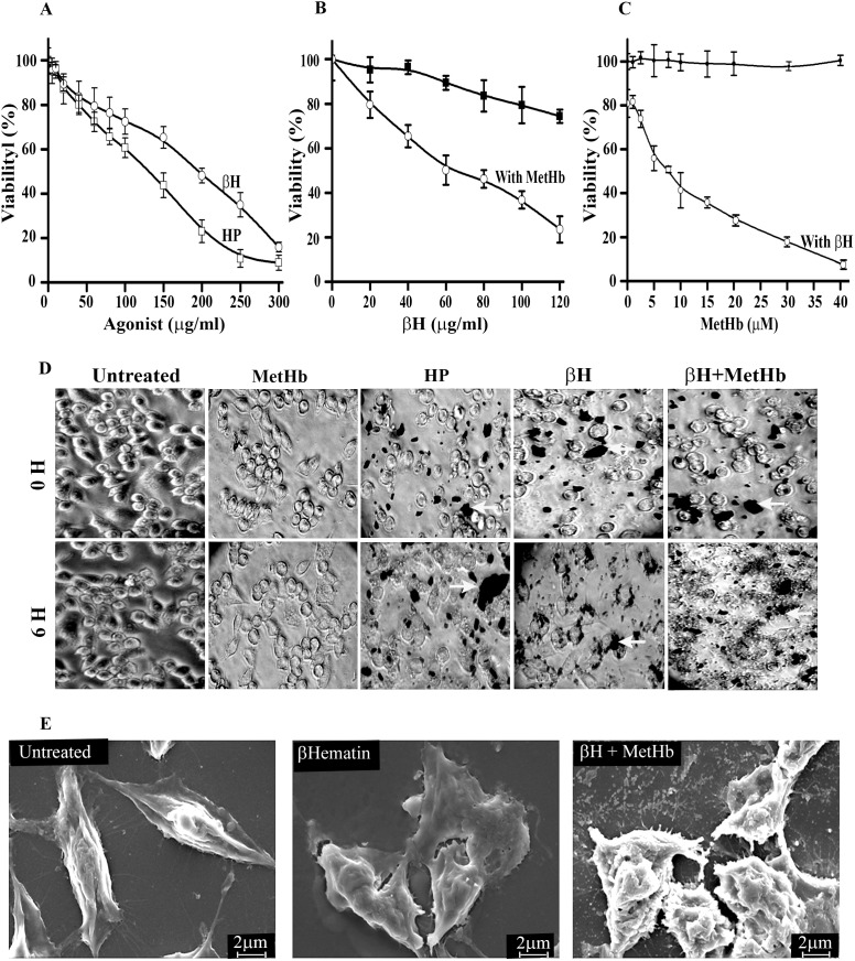
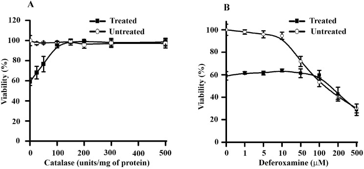
Apoptosis in macrophages is responsible for immune-depression and pathological effects during malaria. Phagocytosis of PRBC causes induction of apoptosis in macrophages through release of cytosolic factors from infected cells. Heme polymer or β-hematin causes dose-dependent death of macrophages with LC50 of 132 µg/ml and 182 µg/ml respectively. The toxicity of hemin or heme polymer was amplified several folds in the presence of non-toxic concentration of methemoglobin. β-hematin uptake in macrophage through phagocytosis is crucial for enhanced toxicological effects in the presence of methemoglobin. Higher accumulation of β-hematin is observed in macrophages treated with β-hematin along with methemoglobin. Light and scanning electron microscopic observations further confirm accumulation of β-hematin with cellular toxicity. Toxicological potentiation of pro-oxidant molecules toward macrophages depends on generation of H2O2 and independent to release of free iron from pro-oxidant molecules. Methemoglobin oxidizes β-hematin to form oxidized β-hematin (βH*) through single electron transfer mechanism. Pre-treatment of reaction mixture with spin-trap Phenyl-N-t-butyl-nitrone dose-dependently reverses the β-hematin toxicity, indicates crucial role of βH* generation with the toxicological potentiation. Acridine orange/ethidium bromide staining and DNA fragmentation analysis indicate that macrophage follows an oxidative stress dependent apoptotic pathway to cause death. In summary, current work highlights mutual co-operation between methemoglobin and different pro-oxidant molecules to enhance toxicity towards macrophages. Hence, methemoglobin peroxidase activity can be probed for subduing cellular toxicity of pro-oxidant molecules and it may in-turn make up for host immune response against the malaria parasite.


Conflict of interest statement

Competing Interests: The authors have declared that no competing interests exist.

Figures

Figure 1
Figure 1. Pro-oxidant molecules co-operate with each other to exhibit enhanced toxicity towards macrophages.
(A) Evaluation of toxicological potential of heme polymer (HP) and β-hematin (βH) against macrophage J774A.1. Macrophages were treated with different concentration of agonist [heme polymer (HP) or β-hematin (βH)] for 6 hr at 37°C and viability was determined by MTT assay as described in “material and methods”. (B) Methemoglobin potentiates the toxicity of β-hematin towards macrophage J774A.1. Macrophages were treated with different concentration of β-hematin (βH) for 6 hr in absence or presence of methemoglobin (7.75 µM) at 37°C and viability was determined by MTT assay as described in “material and methods”. (C) β-hematin potentiates the toxicity of methemoglobin towards macrophage J774A.1. Macrophages were treated with different concentration of methemoglobin (0–40 µM) in absence or presence of β-hematin (60 µg/ml) at 37°C and macrophage viability was determined as described in “material and methods”. In panel (A), (B) and (C), macrophages treated with incomplete media were considerd as 100% viable. Data is the mean ± SD of three independent experiments (n = 3) with triplicate measurement. (D) Microscopic observation of β-hematin particles and cellular damage in macrophages. Macrophages were either untreated or treated with methemoglobin, heme polymer (HP), β-Hematin (βH) or β-Hematin (60 µg/ml)/methemoglobin (7.75 µM) mixture respectively for 6 hr at 37°C and images of random 10 fields were captured with a 20x objective using an invereted microscope TS100 (Nikon, Japan). β-hematin particles are denoted by arrows in each panel. (E) SEM analysis of macrophages. Macrophages were either untreated or treated with β-hematin (60 µg/ml) in the absence or presence of non-toxic concentration of methemoglobin (7.75 µM) for 6 hr at 37°C and a total of 10 different fields were captured using LEO 1430VP Scanning Electron Microscope with the instrument setting as EHT, width and signal were 10 kV, 15 mm and SE1 respectively. A representative image of untreated, treated with β-Hematin (60 µg/ml) or β-hematin (60 µg/ml)/methemoglobin (7.75 µM) mixture respectively (magnification; x2500, scale bar = 2 µm) is given.
Figure 2
Figure 2. Determination of intracellular oxidative stress indices within macrophages.
(A–C) Level of oxidative stress indices in macrophages treated with combination of β-hematin (60 µg/ml)/methemoglobin (7.75 µM) over the course of time. Macrophages were either untreated or treated with the combination of β-hematin (60 µg/ml)/MetHb (7.75 µM) for different time points (0–6 hr) and (A) lipid peroxidation (B) protein carbonyl and (C) reduced glutathione is measured and expressed as mM/mg of cell lysate. Data is the mean ± SD of three independent experiments (n = 3) with triplicate measurement. Cellular viability is measured to correlate the change in oxidative stress with the viability of treated cells. The correlation factor (r2) for change in viability Vs lipid peroxidation is 0.97, change in viability Vs protein carbonyl is 0.86 and change in viability Vs GSH level is 0.97.
Figure 3
Figure 3. Oxidative stress is required for interaction of different pro-oxidant molecules (β-hematin/methemoglobin) to exhibit enhanced toxicity in macrophages.
(A) Removal of oxidative stress through anti-oxidant treatement provides recovery in macrophages from the toxicity of β-hematin/methemoglobin mixture. Macrophages were either untreated or treated with β-hematin (60 µg/ml)/methemoglobin (7.75 µM) for 6 hr at 37°C in the absence or presence of NAC (5 mM) and mannitol (5 mM) respectively. Cell viability was measured by MTT assay as described in “material and methods”. Macrophage treated with incomplete media was considered as 100% viable. Macrophage treated with combination of β-hematin (60 µg/ml)/methemoglobin (7.75 µM) was considered as 0% recovery and the cellular viability in the presence of NAC (5 mM) or mannitol (5 mM) was calculated and expressed as % recovery ±SD. Data is the mean ± SD of three independent experiments (n = 3) with triplicate measurement. The pairwise results were analyzed with Anova & Student t-test and it was considered statistically significant with *P<0.001, #P<0.001. (B) Light microscopic observation of macrophages treated in (A) with 20x objective to detect cellular morphology at 0 hr and 6 hr.
Figure 4
Figure 4. Extracellular H2O2 Generation is responsible for methemoglobin mediated βH toxicological potentiation towards macrophages.
(A) Removal of extracellular H2O2 gives recovery from cytotoxic effects of β-hematin towards macrophages. Macrophages were pre-incubated with different amount of catalase (0–500 U) and either remains untreated or treated with combination of β-hematin (60 µg/ml)/methemoglobin (7.75 µM) for 6 hr at 37°C. Macrophage viability was determined by MTT assay as described in “material and methods” and expressed as % viability ± SD. (B) Scavenging free iron has no effect on reversal of cytotoxic effects of β-hematin towards macrophages. Macrophages were pre-incubated with different amount of deferoxamine (0–500 µM) and either remains untreated or treated with combination of β-hematin (60 µg/ml)/methemoglobin (7.75 µM) for 6 hr at 37°C. Macrophage viability was determined by MTT assay as described in “material and methods” and expressed as % viability ± SD. Data is the mean ± SD of three independent experiments (n = 3) with triplicate measurement.
Figure 5
Figure 5. MetHb and β-hematin interaction generate single electron containing species (β-hematin*) to exhibit cyto-toxicity towards macrophages.
(A) Optical spectra of β-hematin oxidation by methemoglobin. Soret spectra were recorded in 100 mM Tris-HCl buffer, pH 7.4, in a total volume of 0.8 ml. Soret spectrum (a) of MetHb (1 µM); (b) a + H2O2 (100 µM); (c) b + β-hematin (10 µM). Equal concentration of β-hematin (10 µM) was added in the reference cuvette to correct the absorbance in soret region. (B) Scavenging single electron containing species (β-hematin*) restores cellular viability of macrophages. Cells were treated with different concentration of β-hematin (0–150 µg/ml)/methemoglobin (7.75 µM) mixture in the presence of different concentration of PBN (50–300 µM) or remained untreated. Cellular viability was determined by MTT assay as described in “material and methods”. Cells treated with incomplete media was considered as 100% viable. Data is the mean ± SD of three independent experiments (n = 3) with triplicate measurement. (C) Light microscopic observation of macrophages treated in (B) with 20x objective to detect cellular morphology at 0 hr and 6 hrs. (D) Binding of PBN to the oxidized βH. β-hematin was incubated with the different concentration of PBN (0–600 µM) in the presence of MetHb (7.75 µM), H2O2 and optical spectra were recorded.
Figure 6
Figure 6. Macrophages follow oxidative stress mediated apoptosis to exhibit death.
(A) Macrophages either remained untreated or treated with MetHb (7.75 µM), β-Hematin (60 µg/ml), or combination of β-Hematin (60 µg/ml)/MetHb (7.75 µM) for 6 hrs at 37°C in incomplete media. Cells were stained with acridine orange/Et-Br and analyzed in flow cytometry to characterize healthy, early or late apoptotic, and necrotic cell population. (B) DNA fragmentation analysis of macrophages treated with methemoglobin (7.75 µM), different concentration of β-Hematin (40 or 60 µg/ml) or combination of β-hematin (60 µg/ml)/methemoglobin (7.75 µM) in incomplete media for 6 hrs at 37°C. DNA fragmentation analysis was performed as described in “material and methods”. (C) Oxidative stress is essential for induction of apoptosis during treatment of macrophages with combination of β-hematin/methemoglobin. DNA fragmentation analysis of macrophages either remains untreated or treated with combination of β-hematin (60 µg/ml)/methemoglobin (7.75 µM) for 6 hrs at 37°C in the absence or presence of different antioxidant molecules (NAC/mannitol). DNA fragmentation analysis was performed as described in “material and methods”. (D) Methemoglobin mediated generation of βH* in the presence of extracellular H2O2 is responsible for macrophage apoptosis. DNA fragmentation analysis of macrophage remains untreated or treated with combination of β-hematin (60 µg/ml)/methemoglobin (7.75 µM) for 6 hrs at 37°C in absence or presence of catalase (200 units) or PBN (300 µM) respectively. DNA fragmentation analysis was performed as described in “material and methods”. (E) Schematic Diagram of methemoglobin mediated β-hematin toxicological potentiation towards macrophages. A details description is given in the text.

References

    1. Sachs J, Malaney P (2002) The economic and social burden of malaria. Nature 415: 680–685. - PubMed
    1. Hay SI, Okiro EA, Gething PW, Patil AP, Tatem AJ, et al. (2010) Estimating the global clinical burden of Plasmodium falciparum malaria in 2007. PLoS Med 7: e1000290. - PMC - PubMed
    1. Cai H, Kuang R, Gu J, Wang Y (2011) Proteases in malaria parasites - a phylogenomic perspective. Curr Genomics 12: 417–427. - PMC - PubMed
    1. Francis SE, Gluzman IY, Oksman A, Knickerbocker A, Mueller R, et al. (1994) Molecular characterization and inhibition of a Plasmodium falciparum aspartic hemoglobinase. EMBO J 13: 306–317. - PMC - PubMed
    1. Pamplona A, Hanscheid T, Epiphanio S, Mota MM, Vigario AM (2009) Cerebral malaria and the hemolysis/methemoglobin/heme hypothesis: shedding new light on an old disease. Int J Biochem Cell Biol 41: 711–716. - PubMed

MeSH terms

LinkOut - more resources