Review
. 2015 May 7;10(5):852-62.
doi: 10.2215/CJN.10741013. Epub 2014 Jul 30.

Osmotic homeostasis

Affiliations
Review

Osmotic homeostasis

John Danziger et al. Clin J Am Soc Nephrol. .

Erratum in

  • Correction.
    [No authors listed] [No authors listed] Clin J Am Soc Nephrol. 2015 Sep 4;10(9):1703. doi: 10.2215/CJN.08340815. Epub 2015 Aug 19. Clin J Am Soc Nephrol. 2015. PMID: 26289317 Free PMC article. No abstract available.

Abstract

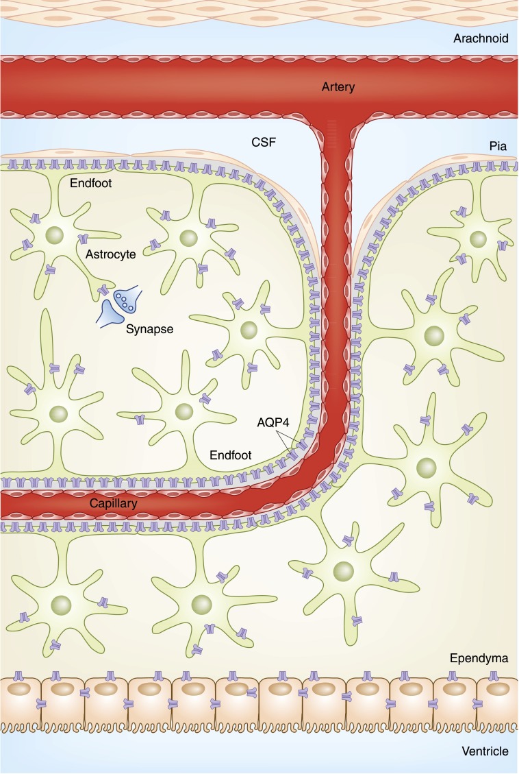
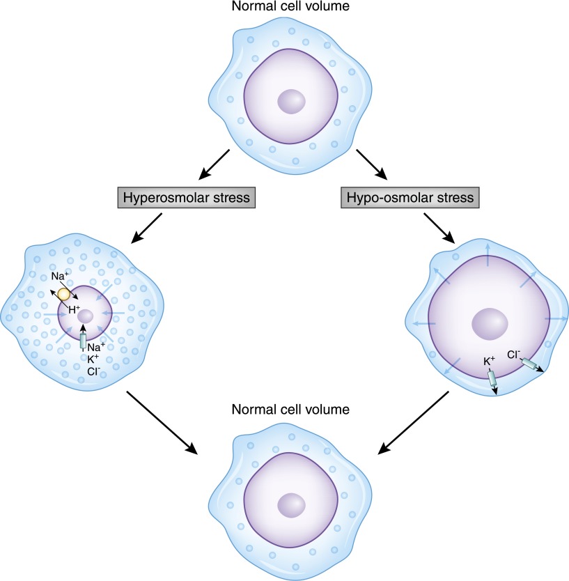
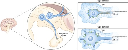
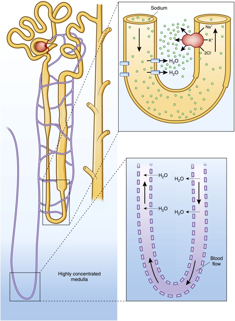
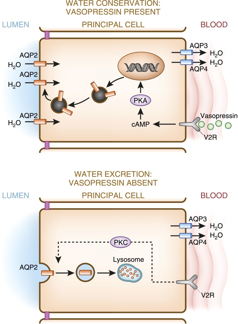
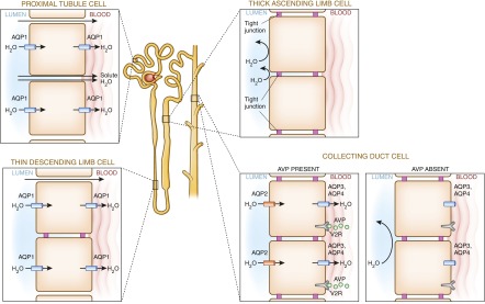
Alterations in water homeostasis can disturb cell size and function. Although most cells can internally regulate cell volume in response to osmolar stress, neurons are particularly at risk given a combination of complex cell function and space restriction within the calvarium. Thus, regulating water balance is fundamental to survival. Through specialized neuronal "osmoreceptors" that sense changes in plasma osmolality, vasopressin release and thirst are titrated in order to achieve water balance. Fine-tuning of water absorption occurs along the collecting duct, and depends on unique structural modifications of renal tubular epithelium that confer a wide range of water permeability. In this article, we review the mechanisms that ensure water homeostasis as well as the fundamentals of disorders of water balance.

Keywords: hypernatremia; hyponatremia; renal physiology; water-electrolyte balance.


Figures

Figure 1.
Figure 1.
The blood-brain barrier. Penetrating capillaries descend through the subarachnoid space into the parenchyma, and are encased by astrocytes, which in addition to controlling important neurologic functions, form the blood-brain barrier. AQP4 water channels along the perivascular and subpial endfoot membranes confer water permeability to the blood-brain barrier. AQP, aquaporin; CSF, cerebrospinal fluid.
Figure 2.
Figure 2.
Cells regulate their internal volume in response to osmotic stress by activation of membrane carrier proteins and channels. In this figure, a normal cell is challenged by either a hyperosmolar (left) or hypo-osmolar (right) milieu. In the setting of hyperosmolar stress, whereby the cell shrinks with water egress, neurons then respond by rapidly accumulating Na+, K+, and Cl ions, followed by the production of intracellular organic solutes. The increase of intracellular solute content then draws water in to normalize the concentrations across the cell membrane, thereby restoring cell size. In the setting of hypo-osmolar–induced swelling, activation of K+ and Cl channels, as well as the K+-Cl cotransporter, lead to solute and consequent water loss, thereby restoring cell volume.
Figure 3.
Figure 3.
Osmoreceptor functions of the OVLT nuclei and SON control thirst and vasopressin release, respectively. In response to hyperosmolar-induced cell shrinkage, specialized mechanical-stretch TRPV cation channels are activated, allowing the influx of positive charges and consequent cell depolarization, provoking action potentials that stimulate thirst and vasopressin release. Conversely, hypo-osmolar cell swelling deactivates these channels, leading to cell hyperpolarization, extinguishing thirst and vasopressin release. Although the exact role of the TRPV channel remains under investigation, its presence is critical in this mechanism. OVLT, organum vasculosum laminae terminalis; SON, supraoptic nuclei; TRPV, transient receptor potential vanilloid.
Figure 4.
Figure 4.
The medullary interstitium has a concentration >4 times that of its surrounding fluid, and must be both generated and maintained. The countercurrent multiplier, composed of a hairpin tubule loop with a water-permeable descending limb juxtaposed against an impermeable ascending limb with a highly active Na-K-2Cl pump, generates the concentration gradient. A separate hairpin loop within the tubular capillary system allows shunting of water from the descending limb to the ascending limb preventing the dilution of the medullary gradient. This process, countercurrent exchange, maintains the medullary concentration.
Figure 5.
Figure 5.
Vasopressin regulates AQP2 expression. In the presence of vasopressin, increased production of cAMP activates PKA, which in turn phosphorylates stored AQP-containing vesicles, and targets them to the apical membrane, increasing its water permeability, and facilitating water reclamation from the lumen. In the absence of vasopressin, AQP2 is endocytosed and internally degraded, conferring water impermeability to the apical membrane, thereby maximizing water excretion. AQP3 and AQP4, constitutively expressed on the basolateral membrane, allow water egress from the cell. PKA, protein kinase A; V2R, vasopressin 2 receptor.
Figure 6.
Figure 6.
Water permeability along the tubule is determined by the presence or absence of intracellular tight junctions and AQP water channels. AQP1, along the proximal tubule and thin descending limb, is constitutively expressed, whereas AQP2, in the collecting duct, is under the control of vasopressin. The presence of AQP1 and the absence of tight junctions render the proximal tubule permeable, facilitating filtered solute and water reclamation (91). In the thin descending limb, the presence of AQP1 and tight junctions (claudin 2) render it water permeable but solute impermeable (92). Conversely, the impermeability of the thick ascending limb results from extensive tight junctions and absent AQP channels. The collecting duct is unique in its homeostatic responsiveness. In times of water conservation, vasopressin (AVP) binds to vasopressin 2 receptors (V2R), inducing AQP2 channel expression and consequent water retention, and in times of water excess, AQP2 retreats from the apical membrane due to vasopressin’s absence.

References

    1. Pitts RF: Physiology of the Kidney and Body Fluids, Chicago, Year Book Medical Publishers, 1963
    1. Epstein FH: The shark rectal gland: A model for the active transport of chloride. Yale J Biol Med 52: 517–523, 1979 - PMC - PubMed
    1. Silva P, Stoff J, Field M, Fine L, Forrest JN, Epstein FH: Mechanism of active chloride secretion by shark rectal gland: Role of Na-K-ATPase in chloride transport. Am J Physiol 233: F298–F306, 1977 - PubMed
    1. Ortiz RM: Osmoregulation in marine mammals. J Exp Biol 204: 1831–1844, 2001 - PubMed
    1. Crone C, Olesen SP: Electrical resistance of brain microvascular endothelium. Brain Res 241: 49–55, 1982 - PubMed

Publication types