Review
. 2015 Aug 7;10(8):1444-58.
doi: 10.2215/CJN.10311013. Epub 2014 Jul 30.

Urea and Ammonia Metabolism and the Control of Renal Nitrogen Excretion

Affiliations
Review

Urea and Ammonia Metabolism and the Control of Renal Nitrogen Excretion

I David Weiner et al. Clin J Am Soc Nephrol. .

Abstract

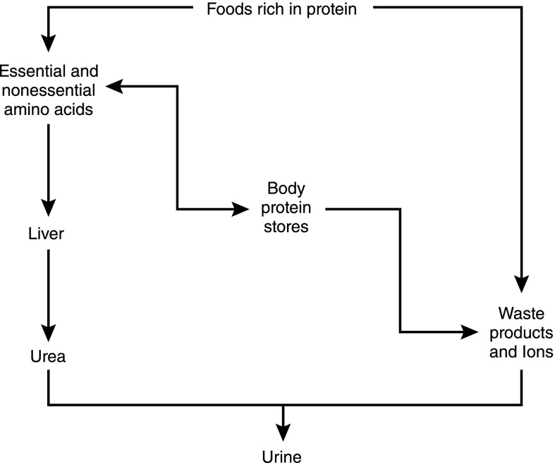
Renal nitrogen metabolism primarily involves urea and ammonia metabolism, and is essential to normal health. Urea is the largest circulating pool of nitrogen, excluding nitrogen in circulating proteins, and its production changes in parallel to the degradation of dietary and endogenous proteins. In addition to serving as a way to excrete nitrogen, urea transport, mediated through specific urea transport proteins, mediates a central role in the urine concentrating mechanism. Renal ammonia excretion, although often considered only in the context of acid-base homeostasis, accounts for approximately 10% of total renal nitrogen excretion under basal conditions, but can increase substantially in a variety of clinical conditions. Because renal ammonia metabolism requires intrarenal ammoniagenesis from glutamine, changes in factors regulating renal ammonia metabolism can have important effects on glutamine in addition to nitrogen balance. This review covers aspects of protein metabolism and the control of the two major molecules involved in renal nitrogen excretion: urea and ammonia. Both urea and ammonia transport can be altered by glucocorticoids and hypokalemia, two conditions that also affect protein metabolism. Clinical conditions associated with altered urine concentrating ability or water homeostasis can result in changes in urea excretion and urea transporters. Clinical conditions associated with altered ammonia excretion can have important effects on nitrogen balance.

Keywords: acidosis; renal physiology; urea.


Figures

Figure 1.
Figure 1.
Overview of protein metabolism. Dietary protein intake can either be metabolized quickly to essential and nonessential amino acids or to metabolic waste products and ions. Essential and nonessential amino acids are interconvertible with body protein stores. Amino acids may also be metabolized through the liver to form urea, which is then excreted in the urine. Body protein stores can be converted back to essential and nonessential amino acids or may be metabolized, forming waste products and ions, which, as previously detailed, are excreted in the urine.
Figure 2.
Figure 2.
Urea transporters along the nephron. The cartoon and histology show the urea transporters (UT-A1/UT-A3, UT-A2, and UT-B1) along the nephron. UT-B1 is found chiefly in the vasa recta, UT-A2 is found in the thin descending limb of the loop of Henle, and UT-A1 (apical) and UT-A3 (basolateral) are found in the inner medullary collecting duct. Modified from reference , with permission.
Figure 3.
Figure 3.
The four renal UT-A protein isoforms. UT-A1 is the largest protein containing 12 transmembrane helices. Helices 6 and 7 are connected by a large intracellular loop that recent studies have shown is crucial to the functional properties of UT-A1 (1). UT-A3 is the N-terminal half of UT-A1, whereas UT-A2 is the C-terminal half of UT-A1. UT-A4 is the N-terminal quarter of UT-A1 spliced to the C-terminal quarter. Modified from reference , with permission.
Figure 4.
Figure 4.
Structure of the nephron. The cartoon depicts the cortex (top), outer medulla (middle), and inner medulla (bottom), showing the location of the various substructures of the nephron labeled as follows: 1, glomerulus; 2, proximal convoluted tubule; 3s and 3l, proximal straight tubule in the short-looped nephron (3s) and long looped nephron (3l); 4s and 4l, thin descending limb; 5, thin ascending limb; 6s and 6l, medullary thick ascending limb; 7, macula densa; 8, distal convoluted tubule; 9, cortical collecting duct; 10, outer medullary collecting duct; 11, initial inner medullary collecting duct; and 12, terminal inner medullary collecting duct. Modified from reference , with permission of the American Physiological Society.
Figure 5.
Figure 5.
Measured urea permeabilities in the different nephron sections of a rat kidney. CCD, cortical collecting duct; DCT, distal convoluted tubule; IMCD, inner medullary collecting duct; mTAL, medullary thick ascending limb; OMCD, outer medullary collecting duct; PCT, proximal convoluted tubule; PST, proximal straight tubule; tAL, thin ascending limb; tDL, thin descending limb. Modified from reference , with permission of the American Physiological Society.
Figure 6.
Figure 6.
Urea transport across an IMCD cell. Vasopressin binds to the V2R, located on the basolateral plasma membrane, and activates the α subunit of the heterotrimeric G protein Gsα. Activation of the G protein stimulates AC to synthesize cAMP. The increase of intracellular cAMP stimulates several downstream proteins including PKA and Epac, which phosphorylate UT-A1 and increase its accumulation in the apical plasma membrane. Urea enters the IMCD cell through UT-A1 and exits on the basolateral plasma membrane via UT-A3. AC, adenylyl cyclase; Epac, exchange protein directly activated by cAMP; Gs, G protein stimulatory subunit; P, phosphate; PKA, protein kinase A; V2R, V2 vasopressin receptor. Modified from reference with permission.
Figure 7.
Figure 7.
Responses of urinary ammonia and titratable acid excretion to exogenous acid loads. Normal humans were acid loaded, and changes in urinary ammonia and titratable acid excretion were determined on days 1, 3, and 5 of acid loading. Changes in urinary ammonia excretion are the quantitatively predominant response mechanism on each day, and continued to increase over the 5 days of the experiment. Titratable acid excretion is a minor component of the increase in net acid excretion, and peaks on day 1 of acid loading. Data calculated from reference .
Figure 8.
Figure 8.
Model of proximal ammonia transport. Glutamine serves as the primary metabolic substrate for ammoniagenesis. Proximal tubule glutamine uptake involves transport across the apical membrane, primarily via BoAT-1, and across the basolateral membrane by SNAT3. Complete metabolism of each glutamine results in generation of two NH4+ and two bicarbonate ions. Bicarbonate is transported across the basolateral membrane via NBCe-1A. Ammonium secretion across the apical membrane occurs primarily via NHE3-mediated Na+/NH4+ exchange, with a lesser contribution by parallel H+ and NH3 transport. BoAT-1, apical Na+-dependent neutral amino acid transporter-1; NBCe-1A, electrogenic sodium-bicarbonate cotransporter, isoform 1A; NHE3, sodium/hydrogen exchanger 3; SNAT3, sodium-coupled neutral amino acid transporter-3.
Figure 9.
Figure 9.
Ammonia reabsorption by the thick ascending limb. Primary mechanism of apical ammonium absorption is via substitution of NH4+ for K+ and transport by the loop diuretic-sensitive, apical NKCC2 transporter. Cytoplasmic NH4+ is transported across the basolateral membrane either via Na+/NH4+ exchange mediated by NHE4 or via a bicarbonate shuttling mechanism involving NH3 transport. NBCn1, electroneutral sodium bicarbonate cotransporter, isoform 1; NHE4, sodium/hydrogen exchanger 4.
Figure 10.
Figure 10.
Ammonia secretion by the collecting duct. Ammonia uptake across the basolateral membrane primarily involves either transporter-mediated uptake across the basolateral membrane by Rhbg or Rhcg, with a component of diffusive NH3 absorption. Cytosolic NH3 is transported across the apical membrane by a combination of Rhcg and diffusive transport. In the IMCD, but not the CCD, basolateral Na+-K+-ATPase also contributes to NH4+ uptake across the basolateral membrane. Cytosolic H+ is generated by a carbonic anhydrase II–mediated mechanism, and is secreted across the apical membrane via H+-ATPase and H+-K+-ATPase. Luminal H+ titrates luminal NH3, forming NH4+ and maintaining a low luminal NH3 concentration necessary for NH3 secretion. CAII, carbonic anhydrase isoform II; Rhbg, Rhesus B glycoprotein; Rhcg, Rhesus C glycoprotein.
Figure 11.
Figure 11.
Integrated overview of renal ammonia metabolism. Renal ammoniagenesis occurs primarily in the proximal tubule, involving glutamine uptake by SNAT3 and BoAT-1, glutamine metabolism forming ammonium and bicarbonate, and apical NH4+ secretion involving NHE3 and parallel H+ and NH3 transport. Ammonia reabsorption in the thick ascending limb, involving apical NKCC2-mediated uptake results in medullary ammonia accumulation. Medullary sulfatides (highlighted in green) reversibly bind NH4+, contributing to medullary accumulation. Ammonia is secreted in the collecting duct via parallel H+ and NH3 secretion. The numbers in blue represent the proportion of total excreted ammonia. BoAT-1, apical Na+-dependent neutral amino acid transporter-1; gsc, galactosylceramide backbone; PDG, phosphate-dependent glutaminase.
Figure 12.
Figure 12.
Urea excretion in adult humans with varying degrees of kidney malfunction fed milk, egg, or an amino acid mixture: assessment of nitrogen balance. Modified from reference , with permission.

References

    1. Mitch WE, Goldberg AL: Mechanisms of muscle wasting. The role of the ubiquitin-proteasome pathway. N Engl J Med 335: 1897–1905, 1996 - PubMed
    1. Lecker SH, Mitch WE: Proteolysis by the ubiquitin-proteasome system and kidney disease. J Am Soc Nephrol 22: 821–824, 2011 - PubMed
    1. Franch HA, Mitch WE: Navigating between the Scylla and Charybdis of prescribing dietary protein for chronic kidney diseases. Annu Rev Nutr 29: 341–364, 2009 - PubMed
    1. Maroni BJ, Steinman TI, Mitch WE: A method for estimating nitrogen intake of patients with chronic renal failure. Kidney Int 27: 58–65, 1985 - PubMed
    1. Addis T: The ratio between the urea content of the urine and of the blood after the administration of large quantities of urea: An approximate index of the quantity of actively functioning kidney tissue. J Urol 1: 263–287, 1917

Publication types