. 2014 Jul;37(7):554-61.
doi: 10.14348/molcells.2014.0159. Epub 2014 Jul 31.

Application of in utero electroporation of G-protein coupled receptor (GPCR) genes, for subcellular localization of hardly identifiable GPCR in mouse cerebral cortex

Affiliations

Application of in utero electroporation of G-protein coupled receptor (GPCR) genes, for subcellular localization of hardly identifiable GPCR in mouse cerebral cortex

Nam-Ho Kim et al. Mol Cells. 2014 Jul.

Abstract

Lysophosphatidic acid (LPA) is a lipid growth factor that exerts diverse biological effects through its cognate receptors (LPA1-LPA6). LPA1, which is predominantly expressed in the brain, plays a pivotal role in brain development. However, the role of LPA1 in neuronal migration has not yet been fully elucidated. Here, we delivered LPA1 to mouse cerebral cortex using in utero electroporation. We demonstrated that neuronal migration in the cerebral cortex was not affected by the overexpression of LPA1. Moreover, these results can be applied to the identification of the localization of LPA1. The subcellular localization of LPA1 was endogenously present in the perinuclear area, and overexpressed LPA1 was located in the plasma membrane. Furthermore, LPA1 in developing mouse cerebral cortex was mainly expressed in the ventricular zone and the cortical plate. In summary, the overexpression of LPA1 did not affect neuronal migration, and the protein expression of LPA1 was mainly located in the ventricular zone and cortical plate within the developing mouse cerebral cortex. These studies have provided information on the role of LPA1 in brain development and on the technical advantages of in utero electroporation.

Keywords: GPCR; cerebral cortex; in utero electroporation; lysophosphatidic acid; lysophosphatidic acid receptor-1.


Figures

Fig. 1.
Fig. 1.
In vivo overexpression of LPA1 protein by in utero electroporation. Confocal images of coronal sections of cortices of embryonic (E13.5) mice transfected by in utero electroporation with control (A, C; expressing EGFP; pCAGIG) or LPA1 (B, D; expressing EGFP and LPA1; pCAGIG-LPA1) constructs that were harvested 48 h post electroporation and immunostained for GFP. Distribution of GFP positive cells in 10 bins spanning cortical thickness are shown as histograms (C, D). DAPI staining was used as a nuclear marker. CP, cortical plate; IZ, intermediate zone; SVZ, subventricular zone; VZ, ventricular zone. Scale bar, 100 μm.
Fig. 2.
Fig. 2.
Western blot analysis of LPA1 proteins in TR neocortical neuroblast cells. TR cells were transiently transfected with control (expressing EGFP) or LPA1 (expressing EGFP and LPA1) constructs. Twenty-four hours post-transfection, the cells were lysed, probed with an anti-LPA1 antibody and a β-actin antibody, and analyzed by Western blots.
Fig. 3.
Fig. 3.
Immunohistochemical localization of LPA1 in the in utero electroporated cortices of embryonic mice. Embryonic mice cortices that were in utero electroporated at E13.5 with control (A-C; expressing EGFP) or LPA1 (E-G; expressing EGFP and LPA1) constructs and harvested at E15.5 were immunostained with anti-GFP antibody (A, E) or anti-LPA1 antibody (B, F). (C) Merge of (A) and (B). (G) Merge of (E) and (F). DAPI staining was used as a nuclear marker. Samples were analyzed by confocal microscopy in the following way: 10 z-stacks of each sample (1 μm apart) were taken and used to generate a two-dimensional image with Zeiss LSM software (Zeiss). Projections of these 2-D images are shown. Scale bar, 20 μm. (D, H) Higher magnification view of the square region in (C) and (G). Scale bar, 10 μm.
Fig. 4.
Fig. 4.
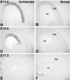
Expression pattern of Lapra1 in mouse embryo. In situ hybridization performed with DIG-labeled riboprobes in the coronal slices showed that Lpar1 transcripts were expressed throughout the ventricular zone at E13.5 (A), E15.5 (C) and E17.5 (E). In situ hybridization with sense probe (B, D, and F) is negative controls. Scale bar, 200 μm.
Fig. 5.
Fig. 5.
Expression pattern of LPA1 in mouse embryo 13.5 cortices. (A) The distribution of LPA1 was examined by immunostaining with an anti-LPA1 antibody (red) in fixed coronal sections of E13.5 embryonic brain. LPA1 protein was expressed in the cells located predominately in the VZ and the lens. (B) Higher magnification view of the square in upper region of A. (B′) Higher magnification view of (B). (C) Higher magnification view of the square in lower region of A. DAPI (blue) staining was used as a nuclear marker. Samples were analyzed by confocal microscopy. (A) Scale bar, 500 μm. (B, C) Scale bar, 50 μm. (B′) Scale bar, 5 μm. Ctx, Cortex; LV, lateral ventricle.
Fig. 6.
Fig. 6.
Expression pattern of LPA1 during cortical development. Sagittal sections of the cortex from E13.5-E17.5 were immunostained to anti-LPA1 antibody (red). DAPI (blue) staining was used as a nuclear marker. Densities of each red signal were measured by dynamic profiler plugin in ImageJ (NIH) and shown in right panel. Samples were analyzed by confocal microscopy. Scale bar, 50 μm. MZ, marginal zone.

References

    1. Aboitiz F, Morales D, Montiel J. The inverted neurogenetic gradient of the mammalian isocortex: development and evolution. Brain Res Brain Res Rev. 2001;38:129–139. - PubMed
    1. An S, Bleu T, Hallmark OG, Goetzl EJ. Characterization of a novel subtype of human G protein-coupled receptor for lysophosphatidic acid. J Biol Chem. 1998;273:7906–7910. - PubMed
    1. Avendano-Vazquez SE, Garcia-Caballero A, Garcia-Sainz JA. Phosphorylation and desensitization of the lysophosphatidic acid receptor LPA1. Biochem J. 2005;385:677–684. - PMC - PubMed
    1. Bandoh K, Aoki J, Hosono H, Kobayashi S, Kobayashi T, Murakami-Murofushi K, Tsujimoto M, Arai H, Inoue K. Molecular cloning and characterization of a novel human G-protein-coupled receptor, EDG7, for lysophosphatidic acid. J Biol Chem. 1999;274:27776–27785. - PubMed
    1. Cheng Y, Makarova N, Tsukahara R, Guo H, Shuyu E, Farrar P, Balazs L, Zhang C, Tigyi G. Lysophosphatidic acid-induced arterial wall remodeling: requirement of PPAR-gamma but not LPA1 or LPA2 GPCR. Cell Signal. 2009;21:1874–1884. - PMC - PubMed

Publication types

MeSH terms

Substances

LinkOut - more resources