Review
. 2014 Sep;82(3):149-62.
doi: 10.5414/cn108386.

Acute interstitial nephritis - a reappraisal and update

Affiliations
Review

Acute interstitial nephritis - a reappraisal and update

Rajeev Raghavan et al. Clin Nephrol. 2014 Sep.

Abstract

Acute interstitial nephritis (AIN) is an under recognized and under diagnosed cause of acute kidney injury (AKI). It is estimated to account for 15 - 20% of cases of AKI; it is the reported diagnosis in 2.8% of all kidney biopsies, and 13.5% of biopsies done specifically for acute renal failure. Considerable evidence implicates antigen initiated cell-mediated injury in the pathogenesis of AIN. Drugs account for 70% of all cases, with over 150 different agents incriminated. The remaining cases are due to infections, autoimmune diseases, and rarely idiopathic. The central component of renal injury in AIN is altered tubular function, which usually precedes decrements in filtration rate. The key to early diagnosis is vigilance for the presence of tubular dysfunction in non-oliguric individuals, especially in patients with modest but gradual increments in creatinine level. The utility of urinary biomarkers to diagnose AIN in its early nascent and potentially reversible stage remains to be determined. Prompt recognition, elimination of the offending source of antigen, and use of a limited course of steroid therapy where indicated, will result in complete resolution in ~ 65% of cases, partial resolution in up to 20%, and irreversible damage in the rest.


Figures

Figure 1.
Figure 1.. Cross section of kidney magnified 5 times to show the foci of regional distribution of cellular infiltrates in acute interstitial nephritis. (Reproduced with permission from reference number [2]).
Figure 2.
Figure 2.. Number of reports listed on PubMed as acute tubulointerstitial nephritis (shown in black) and of acute interstitial nephritis (shown in grey) over the past 45 years. ATIN = acute tubulointerstitial nephritis; AIN = acute interstitial nephritis.
Figure 3.
Figure 3.. Pathogenesis of AIN. The process begins with the recognition and subsequent processing of the putative antigen by dendritic cells that endocytose, process, and express the peptides on their surface MHC II molecules, which they then present to the naïve lymphocytes in the regional lymph nodes. See text under Pathogenesis for the subsequent course of events. EMT = epithelial mesenchymal transition.
Figure 4.
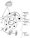
Figure 4.. Schematic representation of the sites of tubular dysfunction in acute tubulointerstitial nephritis. The abnormalities in tubular handling of electrolytes are shown in bold lettering and their clinical manifestation in regular lettering. The boxed black arrows indicate the principal changes reflected in blood and urine tests. PCT = proximal convoluted tubule; DCT = distal convoluted tubule; Loop = loop of Henle; CD = collecting duct; Sp. Gr. = specific gravity; FENa = fractional excretion of sodium; PO4 = phosphate; CO2 = carbon dioxide content. (Reproduced with permission from reference number [6]).

References

    1. Eknoyan G The early modern kidney – nephrology in and about the nineteenth century. Part 1. Semin Dial. 2013; 26: 73–84. - PubMed
    1. Councilman WT Acute interstitial nephritis. J Exp Med. 1898; 3: 393–420. - PMC - PubMed
    1. Johnson G A lecture on scarlet fever and its presentation. BMJ. 1870; 2: 545–546. - PMC - PubMed
    1. Waller BC On the morbid anatomy of certain forms of post-scarlatinal nephritis, in relation to their bearing on the histogeny of granular kidney. J Anat Physiol. 1880; 14: 432–448.. - PMC - PubMed
    1. Biermer MA Ein ungewöhulicher Fall von Scharlach. Virchow’s Arch Pathol Anat. 1860; 19: 537–545.

MeSH terms