

The mammary cellular hierarchy and breast cancer
- PMID: 25080108
- PMCID: PMC4207940
- DOI: 10.1007/s00018-014-1674-4
The mammary cellular hierarchy and breast cancer
Abstract
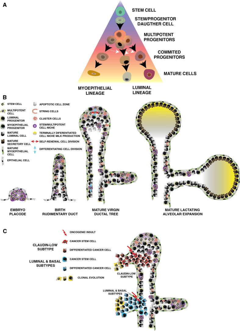
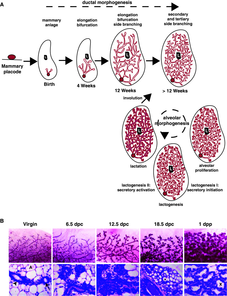
Advances in the study of hematopoietic cell maturation have paved the way to a deeper understanding the stem and progenitor cellular hierarchy in the mammary gland. The mammary epithelium, unlike the hematopoietic cellular hierarchy, sits in a complex niche where communication between epithelial cells and signals from the systemic hormonal milieu, as well as from extra-cellular matrix, influence cell fate decisions and contribute to tissue homeostasis. We review the discovery, definition and regulation of the mammary cellular hierarchy and we describe the development of the concepts that have guided our investigations. We outline recent advances in in vivo lineage tracing that is now challenging many of our assumptions regarding the behavior of mammary stem cells, and we show how understanding these cellular lineages has altered our view of breast cancer.
Figures
Similar articles
-
Mammary gland stem cells and their application in breast cancer.Oncotarget. 2017 Feb 7;8(6):10675-10691. doi: 10.18632/oncotarget.12893. Oncotarget. 2017. PMID: 27793013 Free PMC article. Review.
-
Stem Cells and the Differentiation Hierarchy in Mammary Gland Development.Physiol Rev. 2020 Apr 1;100(2):489-523. doi: 10.1152/physrev.00040.2018. Epub 2019 Sep 20. Physiol Rev. 2020. PMID: 31539305 Review.
-
Delineating the epithelial hierarchy in the mouse mammary gland.Cold Spring Harb Symp Quant Biol. 2008;73:469-78. doi: 10.1101/sqb.2008.73.020. Epub 2008 Nov 6. Cold Spring Harb Symp Quant Biol. 2008. PMID: 19022771 Review.
-
Mammary stem cells and the differentiation hierarchy: current status and perspectives.Genes Dev. 2014 Jun 1;28(11):1143-58. doi: 10.1101/gad.242511.114. Genes Dev. 2014. PMID: 24888586 Free PMC article. Review.
-
The mammary stem cell hierarchy: a looking glass into heterogeneous breast cancer landscapes.Endocr Relat Cancer. 2015 Dec;22(6):T161-76. doi: 10.1530/ERC-15-0263. Epub 2015 Jul 23. Endocr Relat Cancer. 2015. PMID: 26206777 Free PMC article. Review.
Cited by
-
RB1 deficiency in triple-negative breast cancer induces mitochondrial protein translation.J Clin Invest. 2016 Oct 3;126(10):3739-3757. doi: 10.1172/JCI81568. Epub 2016 Aug 29. J Clin Invest. 2016. PMID: 27571409 Free PMC article.
-
The innate and adaptive infiltrating immune systems as targets for breast cancer immunotherapy.Endocr Relat Cancer. 2017 Apr;24(4):R123-R144. doi: 10.1530/ERC-16-0404. Epub 2017 Feb 13. Endocr Relat Cancer. 2017. PMID: 28193698 Free PMC article. Review.
-
Obesity reversibly depletes the basal cell population and enhances mammary epithelial cell estrogen receptor alpha expression and progenitor activity.Breast Cancer Res. 2017 Nov 29;19(1):128. doi: 10.1186/s13058-017-0921-7. Breast Cancer Res. 2017. PMID: 29187227 Free PMC article.
-
Notch Signaling Activation as a Hallmark for Triple-Negative Breast Cancer Subtype.J Oncol. 2019 Jul 11;2019:8707053. doi: 10.1155/2019/8707053. eCollection 2019. J Oncol. 2019. PMID: 31379945 Free PMC article. Review.
-
Evaluation of chemotherapeutic and cancer-protective properties of sphingosine and C2-ceramide in a human breast stem cell derived carcinogenesis model.Int J Oncol. 2019 Feb;54(2):655-664. doi: 10.3892/ijo.2018.4641. Epub 2018 Nov 22. Int J Oncol. 2019. PMID: 30483770 Free PMC article.
References
-
- DeOme KB, et al. Development of mammary tumors from hyperplastic alveolar nodules transplanted into gland-free mammary fat pads of female C3H mice. Cancer Res. 1959;19:515–520. - PubMed
-
- Reya T, et al. Stem cells, cancer, and cancer stem cells. Nature. 2001;414:105–111. - PubMed
-
- Shackleton M, et al. Generation of a functional mammary gland from a single stem cell. Nature. 2006;439:84–88. - PubMed
-
- Stingl J, et al. Purification and unique properties of mammary epithelial stem cells. Nature. 2006;439:993–997. - PubMed
-
- Asselin-Labat ML, et al. Gata-3 is an essential regulator of mammary-gland morphogenesis and luminal-cell differentiation. Nat Cell Biol. 2007;9(2):201–209. - PubMed
Publication types
MeSH terms
Substances
LinkOut - more resources
Full Text Sources
Other Literature Sources
Medical
