Comment
. 2014 Jul 30;5(4):90.
doi: 10.1186/scrt479.

Equine mesenchymal stromal cells and embryo-derived stem cells are immune privileged in vitro

Comment

Equine mesenchymal stromal cells and embryo-derived stem cells are immune privileged in vitro

Yasmin Z Paterson et al. Stem Cell Res Ther. .

Abstract

Introduction: Autologous mesenchymal stem cells (MSCs) are an attractive concept in regenerative medicine, but their mechanism of action remains poorly defined. No immune response is reported after in vivo injection of allogeneic equine MSCs or embryo-derived stem cells (ESCs) into the equine tendon, which may be due to the cells' immune-privileged properties. This study further investigates these properties to determine their potential for clinical application in other tissues.

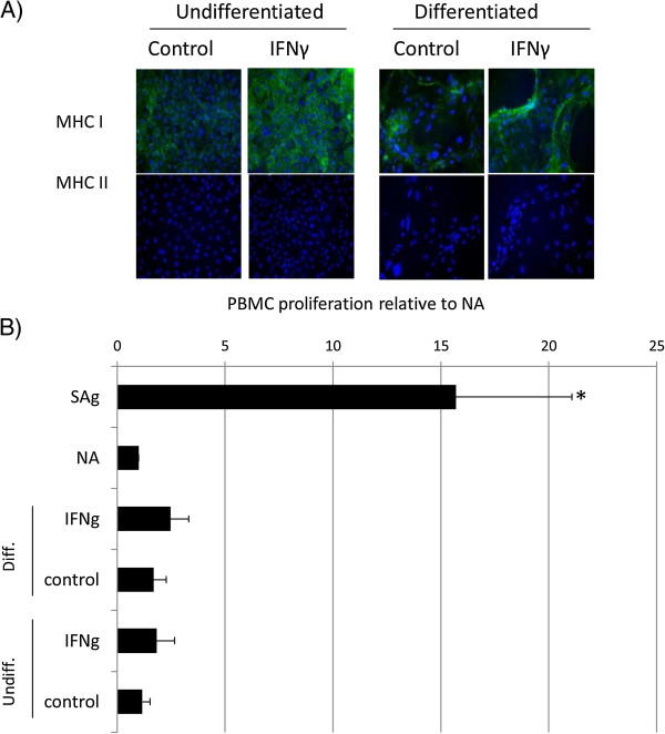
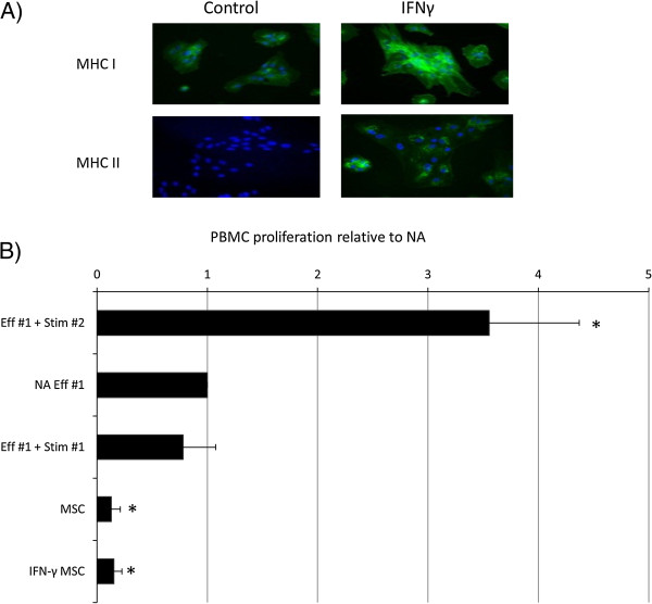
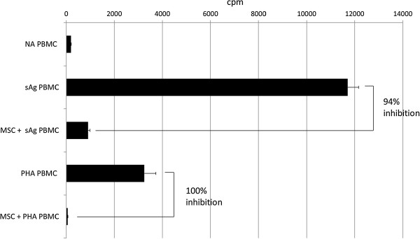
Methods: Mitomycin C-treated MSCs, ESCs, or differentiated ESCs (dESCs) were cultured with allogeneic equine peripheral blood mononuclear cells (PBMCs), and their effect on PBMC proliferation, in the presence or absence of interferon-gamma (IFN-γ) was determined. MSCs and super-antigen (sAg)-stimulated PBMCs were co-cultured directly or indirectly in transwells, and PBMC proliferation examined. Media from MSC culture were harvested and used for PBMC culture; subsequent PBMC proliferation and gene expression were evaluated and media assayed for IFN-γ, tumor necrosis factor alpha (TNF-α), and interleukin (IL)-10 and IL-6 proteins with enzyme-linked immunosorbent assay (ELISA).

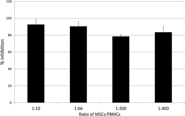
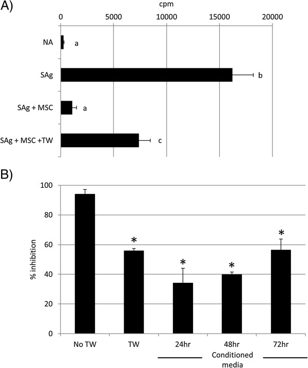
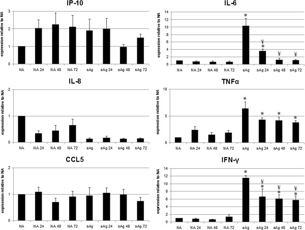
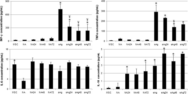
Results: Co-culture of PBMCs with ESCs or dESCs did not affect baseline proliferation, whereas co-culture with MSCs significantly suppressed baseline proliferation. Stimulation of PBMC proliferation by using super-antigens (sAgs) was also suppressed by co-culture with MSCs. Inhibition was greatest with direct contact, but significant inhibition was produced in transwell culture and by using MSC-conditioned media, suggesting that soluble factors play a role in MSC-mediated immune suppression. The MSCs constitutively secrete IL-6, even in the absence of co-culture with PBMCs. MSC-conditioned media also brought about a change in the cytokine-expression profile of sAg-stimulated PBMCs, significantly reducing PBMC expression of IL-6, IFN-γ, and TNF-α.

Conclusions: Equine MSCs and ESCs possess a degree of innate immune privilege, and MSCs secrete soluble factors that suppress PBMC proliferation and alter cytokine expression. These properties may make possible the future clinical use of allogeneic stem cells to help standardize and broaden the scope of treatment of tissue injuries.


Figures

Figure 1
Figure 1
Proliferation of equine PBMCs is not induced by co-culture with equine ESCs. (A) Immunocytochemical staining of IFN-γ-treated embryo-derived stem cells (ESCs) for MHC I and MHC II. Cell nuclei are indicated by blue Dapi staining, and expressed MHC I or II proteins, by green staining. Representative images from one of three replicates are shown. (B) Relative proliferation of peripheral blood mononuclear cells (PBMCs) to undifferentiated (ESCs) and differentiated (dES) ESCs cultured in the presence and absence of IFN-γ, where NA is baseline, nonactivated PBMC proliferation; sAg is superantigen-stimulated PBMCs (positive control); IFN-γ is 72-hour pretreated undifferentiated or differentiated ESC. *Results significantly different relative proliferation when compared with NA PBMCs (P < 0.05). Error bars represent the standard error of seven individual experimental repeats using three different cell lines.
Figure 2
Figure 2
Proliferation of equine PBMCs is suppressed by co-culture with equine MSCs. (A) Immunocytochemical staining of IFN-γ-treated mesenchymal stem cells (MSCs) for MHC I and MHC II. Cell nuclei are indicated by blue Dapi staining, and expressed MHC I or II proteins, by green staining. Data show representative images from one of three replicates. (B) Relative proliferation of effector (Eff#1) peripheral blood mononuclear cells (PBMCs) to mesenchymal stem cells (MSCs) cultured in the presence and absence of IFN-γ. Autologous and allogeneic stimulator cells are used as negative and positive controls (Stim #1 and Stim #2, respectively). *Results significantly different from those for nonactivated (NA) PBMCs (P < 0.05). Error bars represent the standard error of six individual experimental repeats by using three different cell lines.
Figure 3
Figure 3
Proliferation of peripheral blood mononuclear cells (PBMCs) is increased with either S. equi super-antigens (sAg) or phytohemagglutinin (PHA) compared with nonactivated (NA) PBMCs and is inhibited by co-culture with allogeneic mesenchymal stem cells (MSCs). Graph depicts radioactive thymidine (3H-thymidine) counts per minute (cpm) as a measure of cell proliferation. Error bars represent the standard deviation of triplicate wells of the same experiment, which is representative of the three replicates.
Figure 4
Figure 4
Percentage inhibition of S. equi superantigen (sAg)-induced peripheral blood mononuclear cells (PBMCs) proliferation when co-cultured with mesenchymal stem cells (MSCs) at ratios of 1:10, 1:66, 1:200, and 1:400 (MSC to PBMC). Error bars represent the standard error of the mean of three biologic repeats. No significant differences are observed between the ratios (P > 0.05).
Figure 5
Figure 5
Soluble factors are involved in MSC-mediated PMBC suppression. (A) Mesenchymal stem cells (MSCs) suppress S. equi superantigen (sAg)-induced peripheral blood mononuclear cells (PBMCs) proliferation when separated by a transwell membrane (TW). Graph depicts radioactive thymidine (3H-thymidine) counts per minute (cpm) as a measure of cell proliferation. Differing letter annotations denote a significantly different mean (ANOVA, all P < 0.05). Error bars represent the standard error of the mean of three biologic repeats. (B) Exposure to 24-, 48-, and 72-hour mesenchymal stem cell (MSC)-conditioned media suppresses S. equi superantigen (sAg)-induced peripheral blood mononuclear cell (PBMC) proliferation, but to a lesser extent than via direct cell-to-cell contact. TW, transwell. *Results significantly different from no transwell (No TW) values (ANOVA; P < 0.05). Error bars represent the standard error of the mean of three biologic repeats.
Figure 6
Figure 6
mRNA expression by nonactivated (NA) and S. equi superantigens (sAg)-stimulated peripheral blood mononuclear cells (PBMCs) after exposure to 24-, 48-, and 72-hour MSC conditioned media (that is, NA24 represents nonactivated PBMCs exposed to 24-hour MSC-conditioned media). Results represented as change in fold expression relative to NA PBMCs (control conditions), adjusted to the housekeeping gene 18S. Error bars represent the standard error of the mean from three individual experiments using three cell lines. *Results significantly different (P < 0.05) from those for NA PBMCs, ¥ denotes values significantly different (P < 0.05) from sAg-stimulated PBMCs (sAg, stimulated control).
Figure 7
Figure 7
Concentration of cytokines produced by nonactivated (NA) and S. equi superantigen (sAg)-stimulated peripheral blood mononuclear cells (PBMCs) after exposure to 24-, 48-, and 72-hour MSC-conditioned media, measured with ELISA (that is, NA24 represents nonactivated PBMCs exposed to 24-hour MSC-conditioned media). Error bars represent the standard error of the mean from three individual experiments using three cell lines. *Results significantly different from those for nonactivated PBMCs (NA), ¥, Values significantly different (P < 0.05) from sAg-stimulated PBMCs (sAg).

Comment on

References

    1. Smith RK, Korda M, Blunn GW, Goodship AE. Isolation and implantation of autologous equine mesenchymal stem cells from bone marrow into the superficial digital flexor tendon as a potential novel treatment. Equine Vet J. 2003;35:99–102. doi: 10.2746/042516403775467388. - DOI - PubMed
    1. Ouyang HW, Goh JC, Thambyah A, Teoh SH, Lee EH. Knitted poly-lactide-co-glycolide scaffold loaded with bone marrow stromal cells in repair and regeneration of rabbit Achilles tendon. Tissue Eng. 2003;9:431–439. doi: 10.1089/107632703322066615. - DOI - PubMed
    1. Hankemeier S, Keus M, Zeichen J, Jagodzinski M, Barkhausen T, Bosch U, Krettek C, Van Griensven M. Modulation of proliferation and differentiation of human bone marrow stromal cells by fibroblast growth factor 2: potential implications for tissue engineering of tendons and ligaments. Tissue Eng. 2005;11:41–49. doi: 10.1089/ten.2005.11.41. - DOI - PubMed
    1. Godwin EE, Young NJ, Dudhia J, Beamish IC, Smith RK. Implantation of bone marrow-derived mesenchymal stem cells demonstrates improved outcome in horses with overstrain injury of the superficial digital flexor tendon. Equine Vet J. 2012;44:25–32. doi: 10.1111/j.2042-3306.2011.00363.x. - DOI - PubMed
    1. Lange-Consiglio A, Rossi D, Tassan S, Perego R, Cremonesi F, Parolini O. Conditioned medium from horse amniotic membrane-derived multipotent progenitor cells: immunomodulatory activity in vitro and first clinical application in tendon and ligament injuries in vivo. Stem Cells Dev. 2013;22:3015–3024. doi: 10.1089/scd.2013.0214. - DOI - PubMed

Publication types