The probability of seizures during EEG monitoring in critically ill adults
- PMID: 25082090
- PMCID: PMC4289643
- DOI: 10.1016/j.clinph.2014.05.037
The probability of seizures during EEG monitoring in critically ill adults
Abstract
Objective: To characterize the risk for seizures over time in relation to EEG findings in hospitalized adults undergoing continuous EEG monitoring (cEEG).
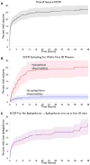
Methods: Retrospective analysis of cEEG data and medical records from 625 consecutive adult inpatients monitored at a tertiary medical center. Using survival analysis methods, we estimated the time-dependent probability that a seizure will occur within the next 72-h, if no seizure has occurred yet, as a function of EEG abnormalities detected so far.
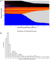
Results: Seizures occurred in 27% (168/625). The first seizure occurred early (<30min of monitoring) in 58% (98/168). In 527 patients without early seizures, 159 (30%) had early epileptiform abnormalities, versus 368 (70%) without. Seizures were eventually detected in 25% of patients with early epileptiform discharges, versus 8% without early discharges. The 72-h risk of seizures declined below 5% if no epileptiform abnormalities were present in the first two hours, whereas 16h of monitoring were required when epileptiform discharges were present. 20% (74/388) of patients without early epileptiform abnormalities later developed them; 23% (17/74) of these ultimately had seizures. Only 4% (12/294) experienced a seizure without preceding epileptiform abnormalities.
Conclusions: Seizure risk in acute neurological illness decays rapidly, at a rate dependent on abnormalities detected early during monitoring. This study demonstrates that substantial risk stratification is possible based on early EEG abnormalities.
Significance: These findings have implications for patient-specific determination of the required duration of cEEG monitoring in hospitalized patients.
Keywords: Continuous electroencephalography; ICU EEG monitoring; Nonconvulsive seizures.
Copyright © 2014 International Federation of Clinical Neurophysiology. Published by Elsevier Ireland Ltd. All rights reserved.
Conflict of interest statement
None of the authors has any conflict of interest to disclose. We confirm that we have read the Journal’s position on issues involved in ethical publication and affirm that this report is consistent with those guidelines.
Figures


Comment in
-
Early epileptiform discharges and the yield of prolonged EEG monitoring.Clin Neurophysiol. 2015 Mar;126(3):431-2. doi: 10.1016/j.clinph.2014.07.002. Epub 2014 Jul 15. Clin Neurophysiol. 2015. PMID: 25127706 No abstract available.
References
-
- Annegers JF, Grabow JD, Groover RV, Laws ER, Jr, Elveback LR, Kurland LT. Seizures after head trauma: a population study. Neurology. 1980;30:683–9. - PubMed
-
- Baker CJ, Prestigiacomo CJ, Solomon RA. Short-term perioperative anticonvulsant prophylaxis for the surgical treatment of low-risk patients with intracranial aneurysms. Neurosurgery. 1995;37:863–70. discussion 870–871. - PubMed
-
- Butzkueven H, Evans AH, Pitman A, Leopold C, Jolley DJ, Kaye AH, et al. Onset seizures independently predict poor outcome after subarachnoid hemorrhage. Neurology. 2000;55:1315–20. - PubMed
-
- Carrera E, Claassen J, Oddo M, Emerson RG, Mayer SA, Hirsch LJ. Continuous electroencephalographic monitoring in critically ill patients with central nervous system infections. Arch Neurol. 2008;65:1612–8. - PubMed
MeSH terms
Grants and funding
LinkOut - more resources
Full Text Sources
Other Literature Sources
Medical

