

Insights into synaptic function from mouse models of human cognitive disorders
- PMID: 25083141
- PMCID: PMC4114080
- DOI: 10.2217/fnl.10.80
Insights into synaptic function from mouse models of human cognitive disorders
Abstract
Modern approaches to the investigation of the molecular mechanisms underlying human cognitive disease often include multidisciplinary examination of animal models engineered with specific mutations that spatially and temporally restrict expression of a gene of interest. This approach not only makes possible the development of animal models that demonstrate phenotypic similarities to their respective human disorders, but has also allowed for significant progress towards understanding the processes that mediate synaptic function and memory formation in the nondiseased state. Examples of successful mouse models where genetic manipulation of the mouse resulted in recapitulation of the symptomatology of the human disorder and was used to significantly expand our understanding of the molecular mechanisms underlying normal synaptic plasticity and memory formation are discussed in this article. These studies have broadened our knowledge of several signal transduction cascades that function throughout life to mediate synaptic physiology. Defining these events is key for developing therapies to address disorders of cognitive ability.
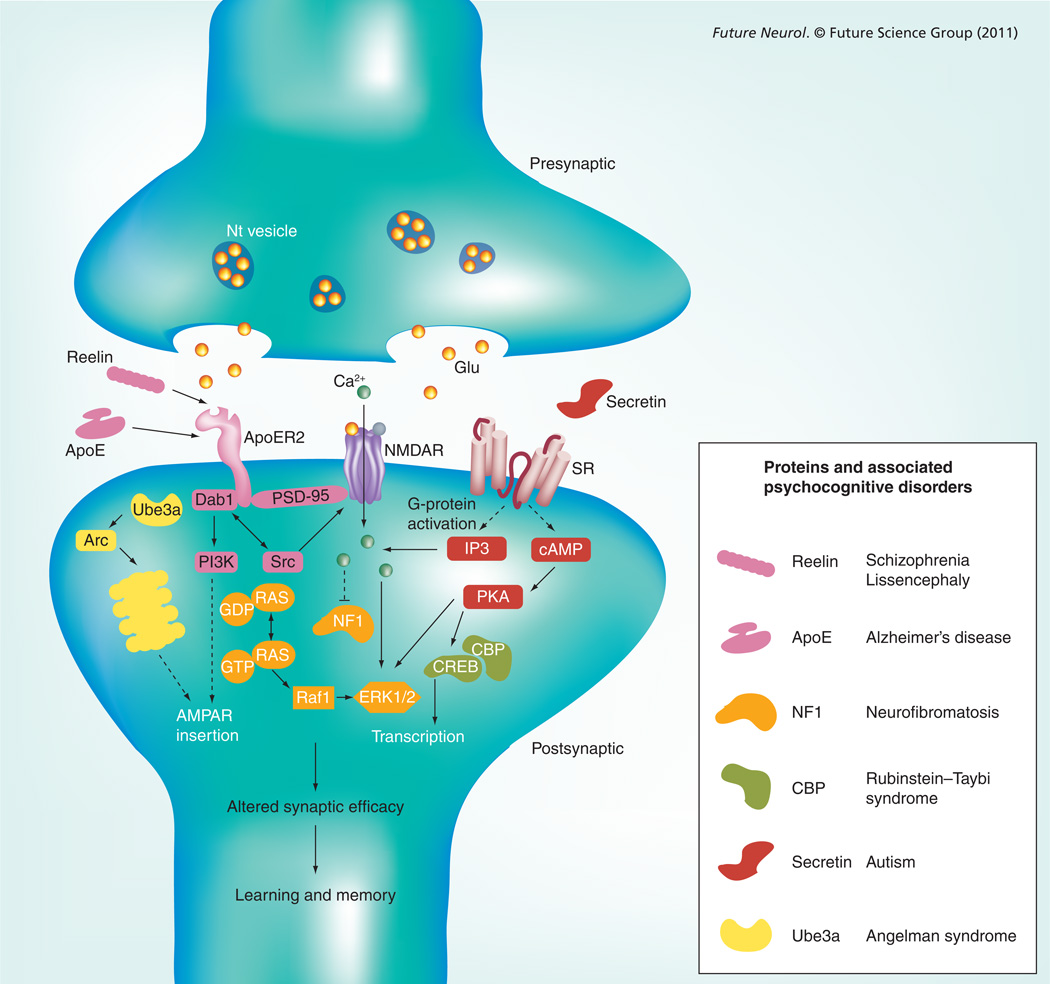
Keywords: Alzheimer’s disease; Angelman syndrome; Reelin; Rubinstein-Taybi syndrome; autism; hippocampus; knockout mouse; neurofibromatosis type 1; secretin; synaptic plasticity.
Conflict of interest statement
The authors have no other relevant affiliations or financial involvement with any organization or entity with a financial interest in or financial conflict with the subject matter or materials discussed in the manuscript apart from those disclosed.
No writing assistance was utilized in the production of this manuscript.
Figures
References
-
- Aboitiz F, Morales D, Montiel J. The evolutionary origin of the mammalian isocortex: towards an integrated developmental and functional approach. Behav. Brain Sci. 2003;26(5):535–552. discussion 52–85. - PubMed
-
- Kerr KM, Agster KL, Furtak SC, Burwell RD. Functional neuroanatomy of the parahippocampal region: the lateral and medial entorhinal areas. Hippocampus. 2007;17(9):697–708. - PubMed
-
- Grover LM, Teyler TJ. Two components of long-term potentiation induced by different patterns of afferent activation. Nature. 1990;347(6292):477–479. - PubMed
Grants and funding
LinkOut - more resources
Full Text Sources
Research Materials