

Optimized contrast-enhanced ultrasonography for characterization of focal liver lesions in cirrhosis: A single-center retrospective study
- PMID: 25083285
- PMCID: PMC4114116
- DOI: 10.1177/2050640614538964
Optimized contrast-enhanced ultrasonography for characterization of focal liver lesions in cirrhosis: A single-center retrospective study
Abstract
Background: Hepatocellular carcinoma (HCC) is the leading cause of death amongst cirrhotic patients. Its diagnosis and discrimination from non-HCC malignant lesions in cirrhosis includes contrast enhanced computed tomography (CECT), contrast enhanced magnetic resonance imaging (CEMRI), or, in selected cases, liver biopsy. The role of contrast-enhanced ultrasonography (CEUS) is still controversial.
Aims: To evaluate whether, by selecting an appropriate 'time to wash-out' cut-off value, CEUS capability of discriminating between HCC and non-HCC malignancies in cirrhotic patients may be enhanced.
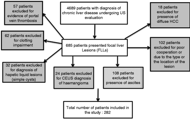
Methods: We enrolled 282 cirrhotic patients who underwent CEUS at our institute, from January 2008 to January 2012, for focal liver lesions (FLLs) detected at ultrasound (US). We used liver biopsy and subsequent histological evaluation as the gold standard for correct classification of FLLs. We calculated the area under receiver operator characteristic curves for CEUS to distinguish patients with HCC from those with non-HCC malignancies. The best 'time to wash-out' cut-off values were selected.
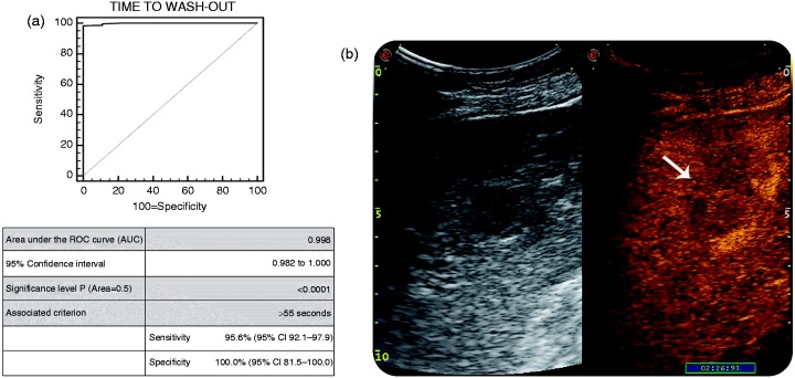
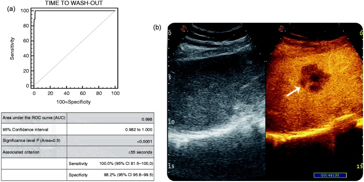
Results: HISTOLOGICAL DIAGNOSIS OF FLLS WAS AS FOLLOWS: 34 benign lesions (i.e. 25 regenerative nodules and 9 dysplastic nodules) and 248 malignant lesions (223 well-to-moderately differentiated HCCs; 7 poorly-differentiated HCCs; 5 intrahepatic colangiocellular carcinomas (ICCs); 5 primary non-Hodgkin B-cell lymphomas (NHBLs); and 8 metastatic liver tumors). A time to wash-out > 55 s identified patients with HCC with the highest level of accuracy (92.7%). Similarly, a time to wash-out ≤ 55 s correctly identified the vast majority of the non-HCC malignancies (100% sensitivity, 98.2% specificity and diagnostic accuracy of 98.3%).
Conclusions: CEUS is an accurate and safe procedure for discriminating FLLs in cirrhotic patients, especially when a cut-off time to wash-out of 55 s is chosen as a reference value.
Keywords: Biopsy; cholangiocellular carcinoma; cirrhosis; contrast enhancement; focal liver lesions; hepatocellular carcinoma; liver; ultrasound.
Figures
References
-
- Llovet JM, Bruix J. Novel advancements in the management of hepatocellular carcinoma in 2008. J Hepatol 2008; 48: S20–S37. - PubMed
-
- Bruix J, Sherman M, Llovet JM, et al. Clinical management of hepatocellular carcinoma. Conclusions of the Barcelona-2000 European Association for the Study of the Liver (EASL) conference. J Hepatol 2001; 35: 421–430. - PubMed
-
- Caturelli E, Bartolucci F, Biasini E, et al. Diagnosis of liver nodules observed in chronic liver disease patients during ultrasound screening for early detection of hepatocellular carcinoma. Am J Gastroenterol 2002; 97: 397–405. - PubMed
-
- Zardi EM, Uwechie V, Picardi A, et al. Liver focal lesions and hepatocellular carcinoma in cirrhotic patients: From screening to diagnosis. Clin Ter 2001; 152: 185–188. - PubMed
LinkOut - more resources
Full Text Sources
Other Literature Sources
Research Materials
