. 2014 Aug 1;9(8):e103620.
doi: 10.1371/journal.pone.0103620. eCollection 2014.

Characterization of the hemocytes in Larvae of Protaetia brevitarsis seulensis: involvement of granulocyte-mediated phagocytosis

Affiliations

Characterization of the hemocytes in Larvae of Protaetia brevitarsis seulensis: involvement of granulocyte-mediated phagocytosis

Hyojung Kwon et al. PLoS One. .

Abstract

Hemocytes are key players in the immune response against pathogens in insects. However, the hemocyte types and their functions in the white-spotted flower chafers, Protaetia brevitarsis seulensis (Kolbe), are not known. In this study, we used various microscopes, molecular probes, and flow cytometric analyses to characterize the hemocytes in P. brevitarsis seulensis. The circulating hemocytes were classified based on their size, morphology, and dye-staining properties into six types, including granulocytes, plasmatocytes, oenocytoids, spherulocytes, prohemocytes, and adipohemocytes. The percentages of circulating hemocyte types were as follows: 13% granulocytes, 20% plasmatocytes, 1% oenocytoids, 5% spherulocytes, 17% prohemocytes, and 44% adipohemocytes. Next, we identified the professional phagocytes, granulocytes, which mediate encapsulation and phagocytosis of pathogens. The granulocytes were immunologically or morphologically activated and phagocytosed potentially hazardous substances in vivo. In addition, we showed that the phagocytosis by granulocytes is associated with autophagy, and that the activation of autophagy could be an efficient way to eliminate pathogens in this system. We also observed a high accumulation of autophagic vacuoles in activated granulocytes, which altered their shape and led to autophagic cell death. Finally, the granulocytes underwent mitotic division thus maintaining their number in vivo.


Conflict of interest statement

Competing Interests: The authors have declared that no competing interests exist.

Figures

Figure 1
Figure 1. Images of hemocytes and average proportional distribution of circulating hemocytes in naïve and challenged larvae.
Confocal images of hemocytes stained with DAPI (blue) for nuclei (N) and with filamentous actin (F-actin; red) for cytoskeleton visualization. Hemocytes were classified as granulocytes (A and G), plasmatocytes (B and H), oenocytoids (C and I), spherulocytes (D and J), prohemocytes (E and K), and adipohemocytes (F and L) on the basis of their size, morphology, and dye-staining properties. Pseudopodia or filopodia on the plasma membrane, are indicated by white arrow (A, B, G, and H). The panels M and N show melanized oenocytoids (M) and granulocyte (N). Hemocyte counting was performed by differential hemocyte count (DHC) and 35 larvae (11,235 hemocytes; 321 hemocytes per larva) were used to determine the percentage of the six circulating hemocyte types; five naïve larvae and three challenged larvae at each time point (4, 8, 12, 24, and 48 h h) post infection. Results are given as means and standard deviation. *(P<0.05). Challenged larvae were infected with E. coli (panel O) or S. cerevisiae (panel P) whereas naïve larvae were injected with sterile saline solution. PR, prohemocytes; PL, plasmatocytes; GR, granulocytes; SP, spherulocytes; OE, oenocytoids; AD, adipohemocytes. Scale bar = 20 µm.
Figure 2
Figure 2. Images of immunologically activated granulocytes and encapsulation.
(A and B) granulocytes changed their shape rapidly and generated large lobopodia-like, fan-like, or amoeba-like structures (indicated by white arrow). The highly polymorphic glittering vacuoles of variable size and round or irregular shape were closely packed in the granulocyte cytoplasm (indicated by red arrow). Although several filopodia were also generated in the plasmatocytes (C; indicated by white arrow), they did not undergo any sudden or rapid changes in shape and had no polymorphic vacuoles in their cytoplasm. With the exception of most granulocytes and some plasmatocytes, the other cell types showed no morphological changes compared to naïve larvae hemocytes (data not shown). (D and E) The granulocytes phagocytosed and engulfed GFP-expressing E. coli at 4 h post infection (GFP-expressing E. coli are indicated by red arrows). (G and H) Phagocytosis of GFP-expressing S. cerevisiae (indicated by red arrow) by GRs (indicated by short white arrow). Encapsulation by granulocytes at 12 h post bacterial (panel F; GFP-expressing E. coli phagocytosed by granulocytes are indicated by white arrows) and yeast infection (panel I; GFP-expressing S. cerevisiae are indicated by red arrow, and granulocytes are indicated by white arrow). GR, granulocytes; PL, plasmatocytes. N, nuclei, Scale bar = 20 µm.
Figure 3
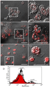
Figure 3. LysoTracker Red labeling of lysosomes in granulocytes and flow cytometric analysis after non-florescence carboxylate-modified polystyrene latex beads injection.
(A and A1) 0 h post injection, (B and B1) 12 h post injection, and (C and C1) 48 h post injection, and (D) flow cytometric analysis at 12 h post injection. Data shows representative histogram overlays (filled histogram, 0 h post injection h post injection; and transparent histogram, 12 h post injection h post injection). The red filled histogram and transparent histogram indicate naïve and challenged larvae, respectively. Based on the red fluorescence intensity, two peaks were identified, Lysolow and Lysohigh. In the Lysolow region, approximately 66.68% of naïve were increased to 77.44% at 12 h post infection. In addition, 1.93 h post infection. In addition, 1.93% of hemocytes of naïve were increased to 7.73% of hemocytes at 12 h post infection in the Lyso h post infection in the Lysohigh region. A1, B1, and C1 indicate a higher magnification of the regions in inset of panel A, B, and C. Over 90% of granulocytes were strongly stained by LysoTracker and the staining was especially prominent in their highly polymorphic vacuoles. GR, granulocytes; N, nuclei, Scale bar = 20 µm.
Figure 4
Figure 4. Complete internalization of large foreign particles by granulocytes.
Hemocytes (average size 20.1 µm, n = 30) from the Stag Beetle Lucanus maculifemoratus (Coleoptera: Lucanidae) were injected into the hemocoel. At 12 h post injection, highly polymorphic glittering vacuoles were generated in the granulocytes. (A through C) The granulocytes engulfed and phagocytosed foreign cells and showed classic structures of phagocytosis (cell-in-cell invasion; a crescent-shaped nuclei) indicated by white arrow. (D through D2) Analysis by confocal microscopy demonstrated a complete internalization of hemocytes from L. maculifemoratus within the granulocytes. In some cases, one, or two, even three foreign hemocytes were phagocytized by the granulocytes (E through E2). D1, D2, E1, and E2 indicate a higher magnification of regions in inset of panel D and E. (F through H) GFP CellTracker dye labeled hemocytes (indicated by white arrow) from L. maculifemoratus were completely internalized by the granulocytes. GR, granulocytes; N, nuclei, Scale bar = 20 µm.
Figure 5
Figure 5. Granulocyte phagocytosis is dependent on foreign particle size.
To examine the granulocyte’s phagocytic competence, human breast cells (MCF10; about two times bigger than granulocytes; average size ∼43 µm, n = 30) were injected into the hemocoel. (A) Autolysed MCF10 cells in the hemocoel; inset shows normal type of MCF10. (B) MCF10 nuclei indicated by red “N”). Shown is encapsulation by granulocytes, which cluster around the MCF10 cell. (C) Exposed nuclei from MCF10 agglomerated together and were phagocytosed by granulocytes; inset, granulocyte amoeba-like structure capturing MCF10 nuclei. (D) MCF10 nuclei (indicated by red “N”) were engulfed and phagocytosed by granulocytes, which showed classic structures of phagocytosis (cell-in-cell invasion; a crescent-shaped nuclei) indicated by black arrow. GR, granulocytes; N (red color), nuclei of MCF10 cells, N (black color), nuclei of granulocytes. Scale bar = 20 µm.
Figure 6
Figure 6. LC3-associated phagocytosis (LAP) by Granulocytes.
Non-florescent carboxyl modified beads were injected into the hemocoel. GFP LC3 was used for detection of autophagosome formation in granulocytes at 0–96 h. (A–G) DIC microscope images and fluorescent microscope images (A1–G1) of granulocytes stained with DAPI and GFP-LC3; and flow cytometric analysis (A2–G2). (H) Confocal images of granulocytes stained with DAPI and GFP LC3 at 24 h post injection. (H1) Representative histogram overlay (filled histogram, 0 h post infection, A2 h post infection, A2; green transparent histogram, 24 h post infection, (E2). Based on the green fluorescence intensity, two peaks were identified, LClow and LChigh. (A2 and E2) 11.40% of granulocytes at 24 h post infection and 1.39 h post infection and 1.39% of granulocytes at 0 h post infection were in the LC h post infection were in the LChigh region, and approximately 10% of GFP-stained hemocytes were increased in the LClow region at 24 h post infection. (F2 and G2) At 48 and 96 h post infection, LC3 accumulation in granulocytes decreased gradually. h post infection, LC3 accumulation in granulocytes decreased gradually.
Figure 7
Figure 7. Flow cytometric analysis of hemocytes stained with Fluorescein isothiocyanate (FITC)-annexin/Propidium Iodide (PI).
Non-florescent carboxylate-modified polystyrene latex beads were injected into the hemocoel at 0, 4, 8, 12, and 48 h. The percentage of PI negative and annexin h. The percentage of PI negative and annexin-V positive hemocytes (LR quadrant) was 0.39, 1.13, 0.00, 0.03, 0.06, and 0.11% at 0, 4, 8, 16, 36, and 72 h post injection, respectively. However, the percentage of PI h post injection, respectively. However, the percentage of PI-positive and annexin-V negative hemocytes (UL quadrant) increased from 6.69% at 0 h to 47.80 h to 47.80% at 72 h post injection. The percentage of PI h post injection. The percentage of PI-positive and annexin-V positive (UR quadrant) was 0.88% at 0 h and 2.25 h and 2.25% at 72 h post injection. h post injection.
Figure 8
Figure 8. Granulocyte mitosis induced by non-florescent carboxyl modified beads injection into the hemocoel.
(A) Analysis by DIC and fluorescent microscopes showed that the granulocytes were mitotically divided at 96 h post injection. (B) Flow cytometric analysis of granulocyte mitosis. Shown are representative histogram overlays: filled histogram (0 h injection h injection) and blue transparent histogram (96 h post injection h post injection). 2.42% of hemocytes in naïve larva were undergoing mitosis, compared to 8.60% of hemocytes in challenged larvae at 96 h post injection. h post injection.

References

    1. Janeway CA, Medzhitov R (2002) Innate immune recognition. Annu Rev Immunol 20: 197–216. - PubMed
    1. Hoffmann JA (2003) The immune response of Drosophila. Nature 426: 33–38. - PubMed
    1. Tsakas S, Marmaras VJ (2010) Insect immunity and its signaling: an overview. Invertebrate Surv J 7: 228–238.
    1. Tauszig S, Emmanuelle J, Hoffmann JA, Imler J (2000) Toll-related receptors and the control of antimicrobial peptide expression in Drosophila. Proc Natl Acad Sci USA 97: 10520–10525. - PMC - PubMed
    1. Silverman N, Maniatis T (2001) NF-κB signaling pathways in mammalian and insect innate immunity. Genes & Dev 15: 2321–2342. - PubMed

Publication types

LinkOut - more resources