. 2014 Jul 31;158(3):607-19.
doi: 10.1016/j.cell.2014.05.047.

MicroRNA directly enhances mitochondrial translation during muscle differentiation

Affiliations

MicroRNA directly enhances mitochondrial translation during muscle differentiation

Xiaorong Zhang et al. Cell. .

Abstract

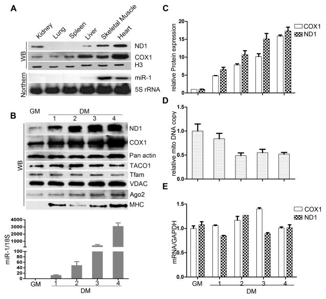
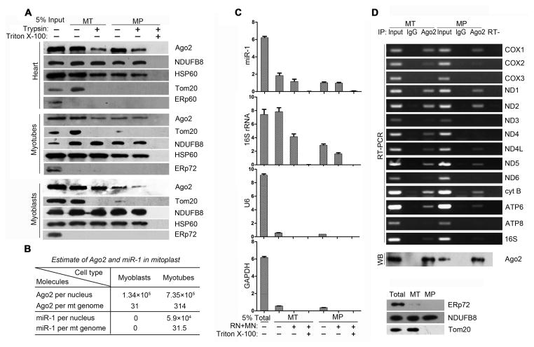
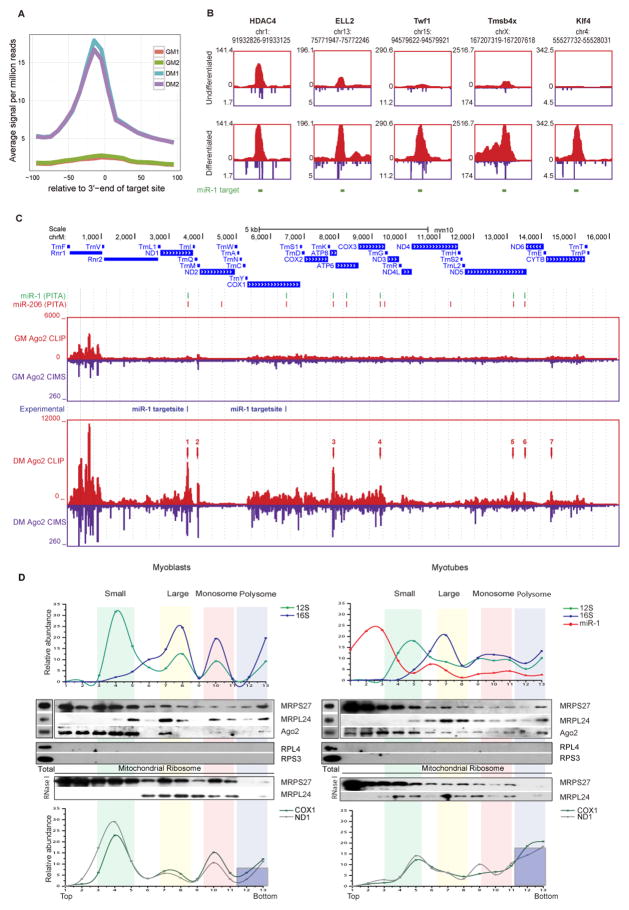
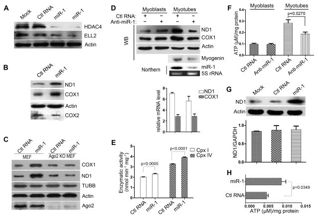
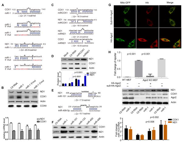
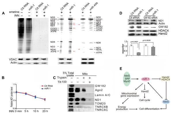
MicroRNAs are well known to mediate translational repression and mRNA degradation in the cytoplasm. Various microRNAs have also been detected in membrane-compartmentalized organelles, but the functional significance has remained elusive. Here, we report that miR-1, a microRNA specifically induced during myogenesis, efficiently enters the mitochondria where it unexpectedly stimulates, rather than represses, the translation of specific mitochondrial genome-encoded transcripts. We show that this positive effect requires specific miR:mRNA base-pairing and Ago2, but not its functional partner GW182, which is excluded from the mitochondria. We provide evidence for the direct action of Ago2 in mitochondrial translation by crosslinking immunoprecipitation coupled with deep sequencing (CLIP-seq), functional rescue with mitochondria-targeted Ago2, and selective inhibition of the microRNA machinery in the cytoplasm. These findings unveil a positive function of microRNA in mitochondrial translation and suggest a highly coordinated myogenic program via miR-1-mediated translational stimulation in the mitochondria and repression in the cytoplasm.


Figures

Figure 1
Figure 1. Increased mitochondrial proteins without a significant elevation in mtDNA copy number or transcription
(A) Levels of COX1 and ND1 proteins detected by Western blotting and miR-1 by Northern blotting in different adult mouse tissues. Levels of Histone H3 and 5S rRNA served as protein and RNA loading controls, respectively. (B) Western and TaqMan PCR analysis of COX1, ND1 and miR-1 in C2C12 cells before and after differentiation. Panactin and VDAC served as protein loading controls and 28S rRNA as an RNA loading control. MHC was analyzed as a control for the induction of the myogenic program. Tfam and TACO1 were analyzed to detect potential regulation of critical mitochondrial transcriptional and translational regulators. (C, D, E) Quantification of proteins normalized to pan actin (C), mtDNA normalized to nuclear DNA (D), and RNA normalized to GAPDH, (E) during C2C12 cell differentiation. See also Fig. S1. Data in C and E are based on three biological repeats and data in D on ten biological repeats. Errors bars are mean ± SD.
Figure 2
Figure 2. Detection and quantitative analysis of Ago2 and miR-1 in the mitochondria
(A) Trypsin protection analysis of purified mitochondria (MT) and mitoplasts (MP) from adult mouse heart, C2C12 myoblasts, and C2C12 myotubes. See also Fig. S2A. (B) Quantification of Ago2 and miR-1 per C2C12 nucleus or per mitochondrial genome. The average number is based on two independent experiments. See also Fig. S2C, D. (C) Nuclease protection analysis. Three aliquots of purified mitochondria and mitoplasts were treated with isolation buffer, with RNaseT1 (RN) plus MNase (MN), or with the combination of nucleases in the presence of Triton X-100. (D) Purified mitochondria and mitoplasts from the same fractionation experiment were first characterized by the lack of the ER marker ERp72, but the presence of outer membrane protein marker Tom20 in the mitochondria and the inner membrane marker NDUFB8 in both mitochondria and mitoplasts (lower panel). Equal amounts of purified mitochondria and mitoplasts based on the levels of 16S rRNA were subjected to ribo-IP with anti-Ago2 or control IgG (upper panel). Western blotting data showed specific anti-Ago2 IP. Individual mitochondrial transcripts were examined by semi-quantitative RT-PCR. See also Fig. S2E, F for real-time RT-PCR analysis of Ago2-associated mitochondrial transcripts from purified mitoplasts.
Figure 3
Figure 3. Mapping of Ago2-RNA interactions by CLIP-seq and analysis of mitochondrial translation by polysome profiling
(A) Ago2 CLIP-seq profiles on predicted miR-1 targets before and after C2C12 differentiation. Duplicated data in each cell type were individually plotted against the center of PITA predicted miR-1 target sites. (B) Ago2 CLIP tags on known cytoplasmic targets for miR-1 before (upper panels) and after (low panels) C2C12 differentiation. The chromosomal location of each target site is shown on the top of each panel. Red: CLIP tags; Blue: CIMS tags. (C) The distribution of Ago2 CLIP-seq tags in the mitochondrial genome before and after C2C12 cell differentiation. The annotated genes are displayed on the top. PITA predicted target sites for miR-1 (green) and miR-206 (red) are indicated right below the gene track. The CLIP tag distribution (red) and corresponding CIMS (dark blue) are separately mapped to the mitochondrial genome in C2C12 myoblasts cultured in growth media (GM) and in C2C12 myotubes maintained in differentiation media (DM) for 4 days. The locations of miRACE identified miR-1 target sites are indicated between the two CLIP panels. (D) Analysis of mitochondrial translation by polysome profiling. Purified mitochondria were used for fractionation on a sucrose gradient, which was free of cytoplasmic ribosomes, as indicated by the lack of Western blotting for representative cytoplasmic ribosomal proteins (RPS3 and RPL4). Representative mitochondrial ribosomal proteins (MRPS27 and MRPL24) and Ago2 on individual gradient fractions were detected by Western blotting and specific rRNA, mRNA and miRNA transcripts were quantified by real-time RT-PCR (Taqman for miR-1). The assignment of small and large ribosomal subunits, monosomes, and putative polysomes was based on the distribution of both rRNAs and proteins and comparison with published mitochondrial polysome profiles. The putative polysome fractions were characterized by RNase I treatment. The relative abundance of individual transcripts in each fraction was presented as the percentage of the total fraction. The increased polysome association of ND1 and COX1 transcripts is highlighted with dark blue bars proportional to their enhancement from myoblasts to myotubes. See also Fig. S3.
Figure 4
Figure 4. Opposite effects of miRNAs on translation in the cytoplasm versus mitochondria
(A) Western blotting of HDAC4 and ELL2 in C2C12 myoblasts after transfection with miR-1 or control siRNA. (B) Western blotting of ND1 and COX1 in C2C12 myoblasts 48 hrs after transfection with control RNA or miR-1. (C) ND1 and COX1 protein levels in wild-type and Ago2 knockout MEFs transfected with either control RNA or miR-1. (D) Antagomir-1 blocked miR-1 expression and diminished enhancedND1 and COX1 translation in myotubes. In this experiment, C2C12 myoblasts were first transfected with control RNA orantagomir-1 and subsequently transferred to differentiation media after 24 hrs. Western blotting of ND1 and COX1 was performed after culturing the cells for another 3 days. Actin and 5S rRNA served as loading controls for protein and RNA. The lower panel showed that antagomir-1 had no effect on the mRNA levels of both ND1 and COX1. (E) Enhanced activities of Complex I and IV by miR-1 in transfected C2C12 myoblasts. (F) Antagomir-1 blocked enhanced ATP production in C2C12 myotubes. (G) miR-1 enhanced ND1 protein synthesis without affect its mRNA levels in transfected HeLa cells. (H) miR-1 induced ATP production in transfected HeLa cells. Data in D, E, F, G, H are based on three biological repeats and shown as mean ± SD; p-values are indicated in each panel. See also Fig. S4.
Figure 5
Figure 5. Target specific effect of miRNA and miRNA mimics
(A) Sequences of miR-1 and its 3′ and 5′ mutants. Calculated free energy for base-pairing is indicated for wild-type miR-1. (B) Analysis of C2C12 myoblasts transfected with wild-type and mutant miR-1 by Western blotting (upper panel) and real time RT-qPCR (bottom panel). Data in the bottom panel are based on triplicated experiments and shown as mean ± SD. (C) Designer miRNA mimics and their base-pairing potential with COX1 and ND1. According to the calculated free energy, miRCOX1 showed reduced base pairing with ND1 whereas miRND1 lost the ability to base pair with COX1. (D) Analysis of C2C12 myoblasts transfected with miRCOX1 and miRND1 by Western (upper panel) and specific proteins levels were quantified (lower panel). (E) Base pairing potentials of a miR-101 and miR-499-5p with distinct regions in the ND1 transcript. (F). Analysis of C2C12 myoblasts transfected with a panel of miRNAs, two of which (miR-101 and miR-499-5p) enhanced ND1, but not COX1, protein synthesis. miR-1 effects were analyzed in parallel as a positive control. See also Fig. S5. (G) Confocal microscopic analysis of Ago2 knockout MEFs complemented with HA-Ago2 or mitochondrial targeted Su9-HA-Ago2. Mitochondria was marked by a co-transfected Mito-GFP. (H) General and selective rescue of Ago2 functions on Ago2 knockout MEFs. A luciferase reporter containing a perfectly complementary sequence to miR-1 was analyzed to the Ago2-mediated RNAi effect in the cytoplasm (upper panel). The data showed that the RNAi effect could be rescued by HA-Ago2, but not mitochondria- targeted Su9-HA-Ago2. The expression of tagged Ago2 and the response of ND1 and COX1 translation of transfected miR-1 in different Ago2 complemented MEFs were shown in the middle panel. The data of triplicated experiments were quantified and shown as mean ± SD with indicated p-values (lower panel).
Figure 6
Figure 6. miR-1 enhanced protein synthesis in the mitochondria
(A) Detection of nascent protein synthesis in the mitochondria when cytoplasmic translation was blocked by Emetine. C2C12 myoblasts were first transfected with a control RNA or miR-1 or miR-499-5p and protein synthesis was monitored by AHA incorporation in the presence of Emetine. Cytoplasmic Actin and mitochondrial VDAC served as loading controls. Emetine blocked all cytoplasmic translation to allow detection of mitochondrial translational activities. Red arrows highlight individual elevated bands by miR-1 or miR-499-5p. (B) Lack of effect of miR-1 on ND1 protein stability. The protein levels were determined in the presence of INN in control and miR-1 transfected C2C12 cells and the data were normalized against the protein level of ND1 at the 0 time point. (C) Restriction of GW182 and its paralogs TNRC6B and TNRC6C from the mitochondria. Lamin A/C was purified away from the mitochondria. ND1 and Tom20 served as controls for proteins localized within the mitochondria or associated with the outer membrane of purified mitochondria. (D) miR-1 enhanced mitochondrial translation when the miRNA machinery was selectively inactivated by knocking down GW182 in the cytoplasm. GW182 showed the expected requirement for miR-1 to act on its cytoplasmic targets HDAC4 and Hand2 (upper panel) and on a cytoplasmic luciferase reporter containing the miR-1 targeting site in ND1 (lower panel). When the function of the miRNA machinery was largely compromised in the cytoplasm of GW182 knockdown cells, transfected miR-1 continued to show the ability to enhance ND1 translation in the mitochondria. (E) Expanded roles of miR-1 in muscle differentiation. miR-1 is known to be induced by SRF, MyoD and Mef2 during differentiation. Induced miR-1 has been thought to act on its cytoplasmic targets to inhibit cell proliferation via Hand2 and promote cell differentiation through HDAC4 and Mef2. The newly elucidated function of miR-1 contributes to efficient protein synthesis in the mitochondria, resulting in boosted ATP production to meet the increasing energy demand in differentiating muscle cells. Therefore, miR-1 mediated translational repression in the cytoplasm and translational enhancement in the mitochondria may constitute a highly coordinated myogenic program for muscle differentiation and function. Data in B and D are based on three biological repeats and shown as mean ± SD with p-values indicated in D.

Comment in

References

    1. Antonicka H, Sasarman F, Nishimura T, Paupe V, Shoubridge EA. The mitochondrial RNA-binding protein GRSF1 localizes to RNA granules and is required for posttranscriptional mitochondrial gene expression. Cell metabolism. 2013;17:386–398. - PubMed
    1. Arnold MA, Kim Y, Czubryt MP, Phan D, McAnally J, Qi X, Shelton JM, Richardson JA, Bassel-Duby R, Olson EN. MEF2C transcription factor controls chondrocyte hypertrophy and bone development. Developmental cell. 2007;12:377–389. - PubMed
    1. Bandiera S, Ruberg S, Girard M, Cagnard N, Hanein S, Chretien D, Munnich A, Lyonnet S, Henrion-Caude A. Nuclear outsourcing of RNA interference components to human mitochondria. PloS one. 2011;6:e20746. - PMC - PubMed
    1. Barrey E, Saint-Auret G, Bonnamy B, Damas D, Boyer O, Gidrol X. Pre-microRNA and mature microRNA in human mitochondria. PloS one. 2011;6:e20220. - PMC - PubMed
    1. Bestwick ML, Shadel GS. Accessorizing the human mitochondrial transcription machinery. Trends in biochemical sciences. 2013;38:283–291. - PMC - PubMed

Publication types

Associated data