

Fundus changes in central retinal vein occlusion
- PMID: 25084156
- PMCID: PMC4276535
- DOI: 10.1097/IAE.0000000000000256
Fundus changes in central retinal vein occlusion
Abstract
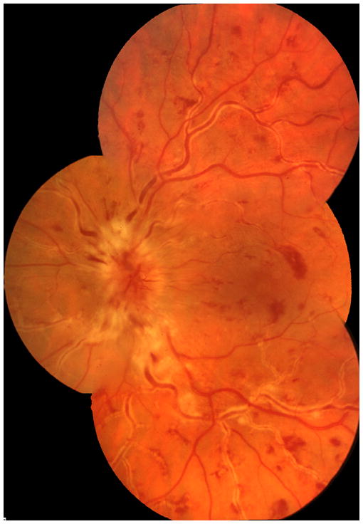
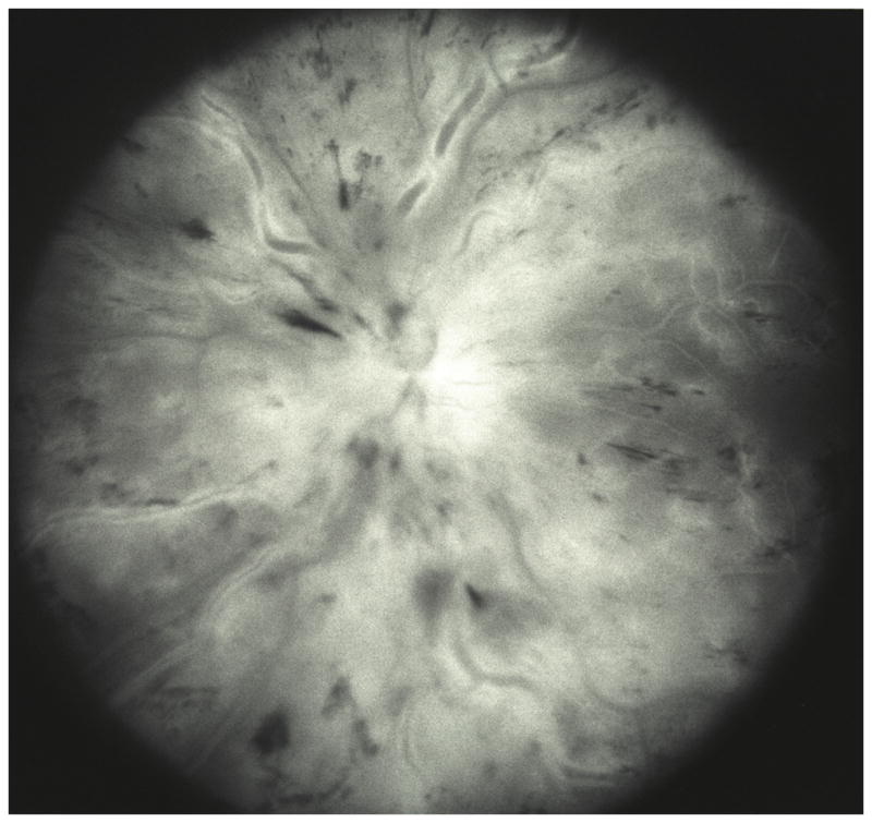
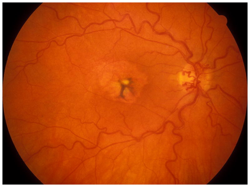
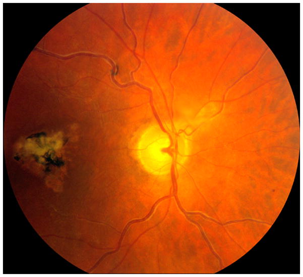
Purpose: To investigate systematically the retinal and optic disk changes in central retinal vein occlusion (CRVO) and their natural history.
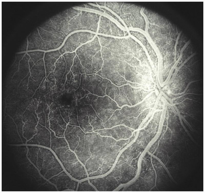
Methods: This study comprised 562 consecutive patients with CRVO (492 nonischemic [NI-CRVO] and 89 ischemic CRVO [I-CRVO] eyes) seen within 3 months of onset. Ophthalmic evaluation at initial and follow-up visits included recording visual acuity, visual fields, and detailed anterior segment and fundus examinations and fluorescein fundus angiography.
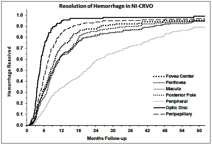
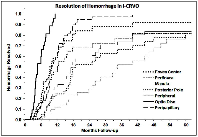
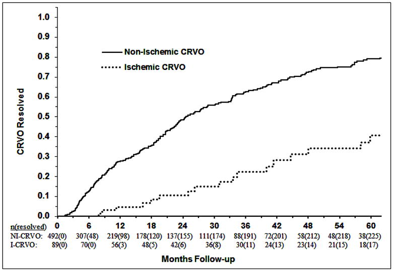
Results: Retinal and subinternal limiting membrane hemorrhages and optic disk edema in I-CRVO were initially more marked (P < 0.0001) and took longer to resolve (P < 0.015) than that in NI-CRVO. Initially, macular edema was more marked in I-CRVO than that in NI-CRVO (P < 0.0001) but did not significantly differ in resolution time (P = 0.238). Macular retinal epithelial pigment degeneration, serous macular detachment, and retinal perivenous sheathing developed at a higher rate in I-CRVO than that in NI-CRVO (P < 0.0001). Ischemic CRVO had more retinal venous engorgement than NI-CRVO (P = 0.003). Fluorescein fundus angiography showed significantly more fluorescein leakage, retinal capillary dilatation, capillary obliteration, and broken capillary foveal arcade (P < 0.0001) in I-CRVO than NI-CRVO. Resolution time of CRVO was longer for I-CRVO than NI-CRVO (P < 0.0001).
Conclusion: Characteristics and natural history of fundus findings in the two types of CRVO are different.
Conflict of interest statement
Authors have no financial interest or conflict.
Figures
References
-
- Michel J. Ueber die anatomischen Ursachen von Veranderungen des Augenhintergrundes bei einigen Allgemeinerkrankungen. Deutsch Arch Klin Med. 1878;22:339–45.
-
- Hayreh SS. So called "central retinal vein occlusion": 1. Pathogenesis, terminology, clinical features. Ophthalmologica. 1976;172:1–13. - PubMed
-
- Hayreh SS. Pathogenesis of occlusion of the central retinal vessels. Am J Ophthalmol. 1971;72:998–1011. - PubMed
-
- Hayreh SS, van Heuven WAJ, Hayreh MS. Experimental retinal vascular occlusion I. Pathogenesis of central retinal vein occlusion. Arch Ophthalmol. 1978;96:311–23. - PubMed
Publication types
MeSH terms
Grants and funding
LinkOut - more resources
Full Text Sources
Other Literature Sources
