Review
. 2014 Aug;94(4):793-815.
doi: 10.1016/j.suc.2014.05.005.

Biology and principles of scar management and burn reconstruction

Affiliations
Review

Biology and principles of scar management and burn reconstruction

Edward E Tredget et al. Surg Clin North Am. 2014 Aug.

Abstract

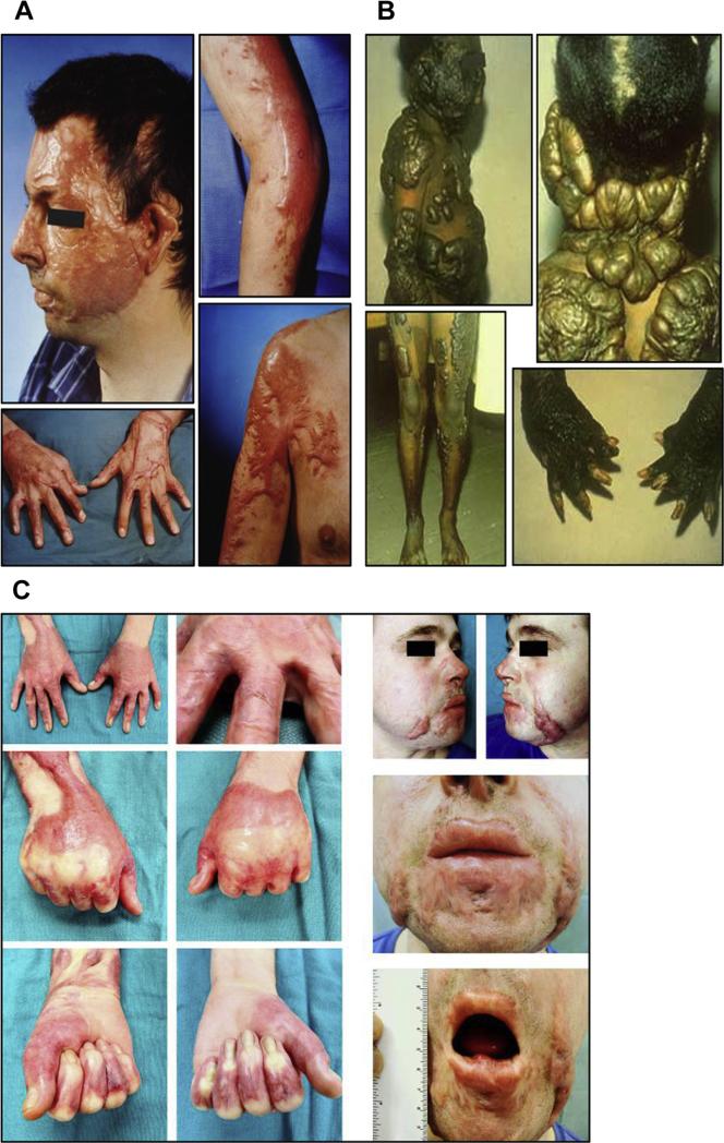
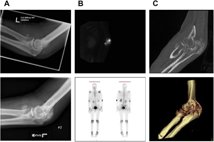
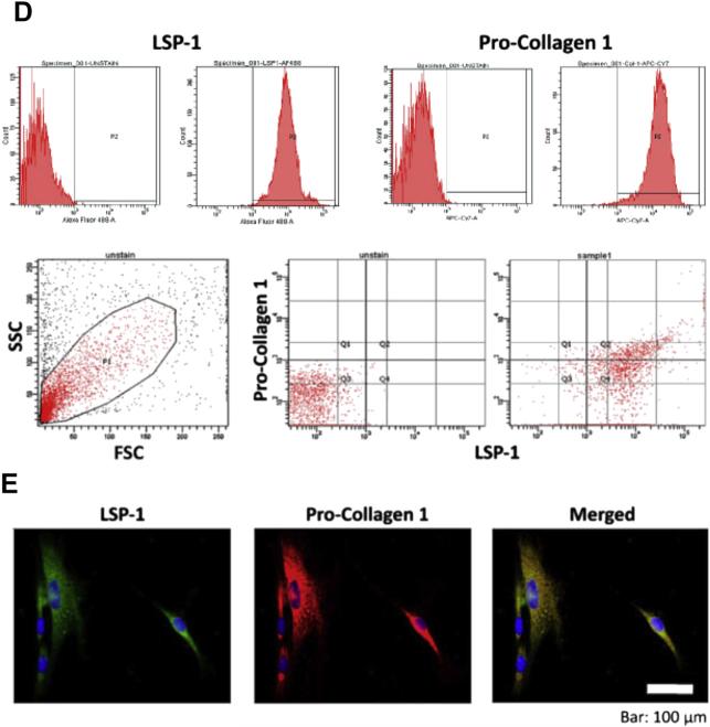
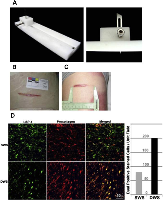
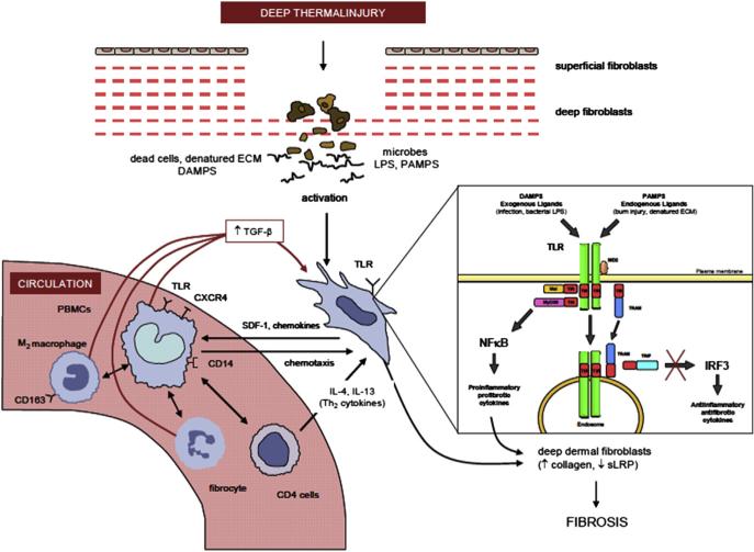
Hypertrophic scarring is extremely common and is the source of most morbidity related to burns. The biology of hypertrophic healing is complex and poorly understood. Multiple host and injury factors contribute, but protracted healing of partial thickness injury is a common theme. Hypertrophic scarring and heterotopic ossification may share some basic causes involving marrow-derived cells. Several traditional clinical interventions exist to modify hypertrophic scar. All have limited efficacy. Laser interventions for scar modification show promise, but as yet do not provide a definitive solution. Their efficacy is only seen when used as part of a multimodality scar management program.

Keywords: Burn reconstruction; Fibrocytes; Heterotopic ossification; Laser; Scar.


Figures

Fig. 1
Fig. 1
(A) HTS developed on a 34-year-old Caucasian man 8 months after a burn involving 60% of his TBSA. (B) Keloids on a 12-year-old black child following a scald burn including donor sites on lower extremities. (C) A 24-year-old white man, 11 months after a 21% TBSA burn. This patient developed HTS, resulting in cosmetic and functional problems that included restricted opening of mouth and tight web spaces of fingers that limited range of motion on hands.
Fig. 2
Fig. 2
A 26-year-old man with 75% TBSA burns who developed HO in both elbows. (A) Imaging studies of the right elbow at 1.5 months (left) and 5 months (right) after burn injury demonstrate the progression of the HO lesion. (B) Intraoperative views from the same patient showing the surgical approach for HO resection (left) and HO specimen (right). (C) Isolation of bone marrow–derived precursor cells from HO tissue by using cell explantation method. A significant cell subset isolated from HO tissue (~35–65%) exhibits a LSP11/COL11 profile as demonstrated by flow cytometry (D) and immune fluorescence microscopy (E).
Fig. 2
Fig. 2
A 26-year-old man with 75% TBSA burns who developed HO in both elbows. (A) Imaging studies of the right elbow at 1.5 months (left) and 5 months (right) after burn injury demonstrate the progression of the HO lesion. (B) Intraoperative views from the same patient showing the surgical approach for HO resection (left) and HO specimen (right). (C) Isolation of bone marrow–derived precursor cells from HO tissue by using cell explantation method. A significant cell subset isolated from HO tissue (~35–65%) exhibits a LSP11/COL11 profile as demonstrated by flow cytometry (D) and immune fluorescence microscopy (E).
Fig. 3
Fig. 3
(A) Jig used to make progressive human dermal scratch injury. (B) Progressive wound on day 0. (C) Wound on day 70. (D) Increased number of fibrocytes in the deeper areas of the wound (DWS) as compared with the superficial wound site (SWS).
Fig. 4
Fig. 4
It is hypothesized that burn injury activates fibroblasts in the deep dermis by using PAMPs (ie, LPS) and DAMPs (ie, Biglycan) to stimulate the Toll receptors/NFκB pathway on fibroblasts, which in turn release chemokines and growth factors (ie, TGF-β) recruiting bone marrow–derived monocytes precursors to further activate the production of ECM proteins in deep dermal fibroblasts and subsequently HTS.
Fig. 5
Fig. 5
Diagramic depiction of the design of various z plastids and their variants with the resultant configuration once cut out and flaps transposed.

References

    1. Kwan P, Desmouliere A, Tredget EE. Chapter 45—Molecular and cellular basis of hypertrophic scarring. In: Herndon DN, editor. Total burn care. 3rd edition. Saunders Elsevier; Philadelphia: 2012. pp. 495–505.e5.
    1. Bombaro KM, Engrav LH, Carrougher GJ, et al. What is the prevalence of hyper-trophic scarring following burns? Burns. 2003;29:299–302. - PubMed
    1. Engrav LH, Covey MH, Dutcher KD, et al. Impairment, time out of school, and time off from work after burns. Plast Reconstr Surg. 1987;79:927–34. - PubMed
    1. Helm P, Herndon DN, Delateur B. Restoration of function. J Burn Care Res. 2007;28(4):611–4. - PubMed
    1. Brown JJ, Bayat A. Genetic susceptibility to raised dermal scarring. Br J Dermatol. 2009;161(1):8–18. - PubMed

Publication types

MeSH terms