. 2014 Nov;96(5):883-94.
doi: 10.1189/jlb.3A0114-012RR. Epub 2014 Aug 1.

Liver metastases induce reversible hepatic B cell dysfunction mediated by Gr-1+CD11b+ myeloid cells

Affiliations

Liver metastases induce reversible hepatic B cell dysfunction mediated by Gr-1+CD11b+ myeloid cells

Mitchell Thorn et al. J Leukoc Biol. 2014 Nov.

Abstract

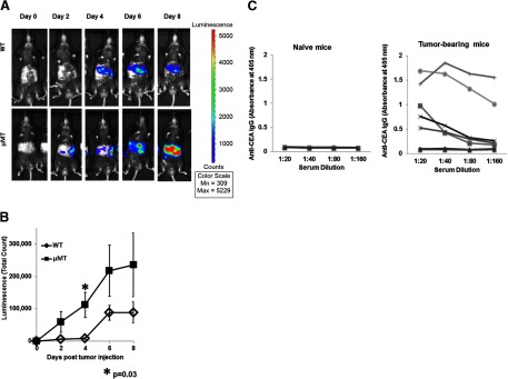
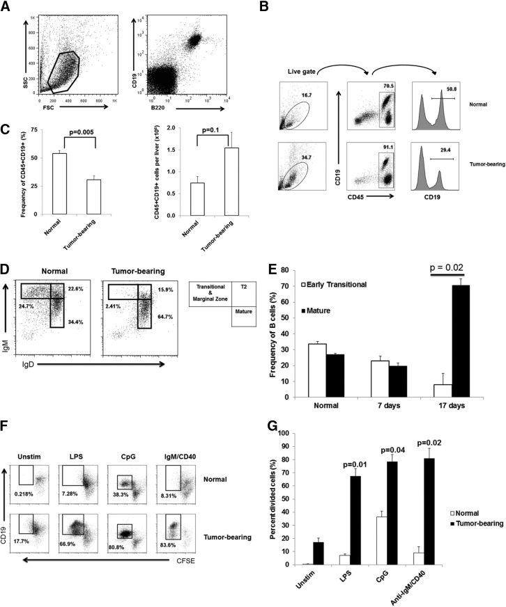
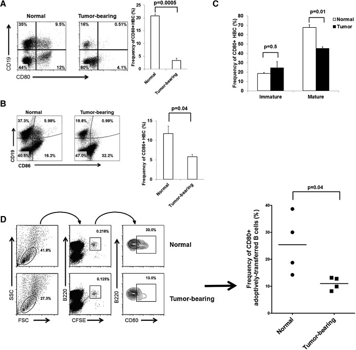
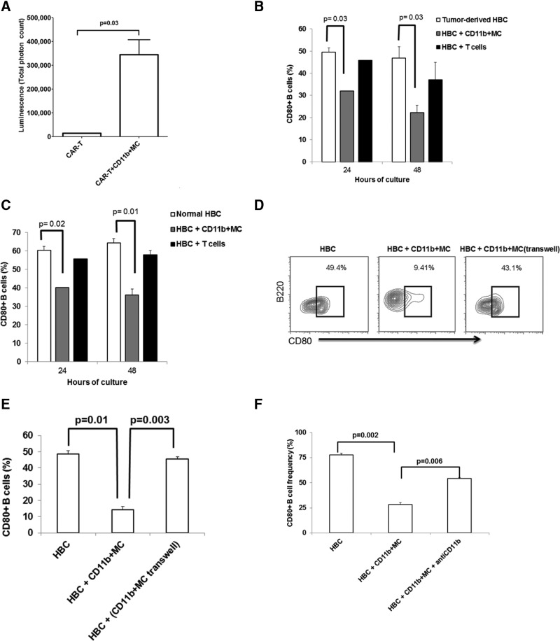
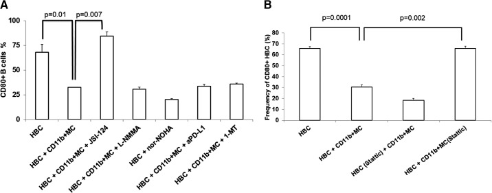
LM escape immune surveillance, in part, as a result of the expansion of CD11b+MC, which alter the intrahepatic microenvironment to promote tumor tolerance. HBC make up a significant proportion of liver lymphocytes and appear to delay tumor progression; however, their significance in the setting of LM is poorly defined. Therefore, we characterized HBC and HBC/CD11b+MC interactions using a murine model of LM. Tumor-bearing livers showed a trend toward elevated absolute numbers of CD19+ HBC. A significant increase in the frequency of IgM(lo)IgD(hi) mature HBC was observed in mice with LM compared with normal mice. HBC derived from tumor-bearing mice demonstrated increased proliferation in response to TLR and BCR stimulation ex vivo compared with HBC from normal livers. HBC from tumor-bearing livers exhibited significant down-regulation of CD80 and were impaired in inducing CD4(+) T cell proliferation ex vivo. We implicated hepatic CD11b+MC as mediators of CD80 down-modulation on HBC ex vivo via a CD11b-dependent mechanism that required cell-to-cell contact and STAT3 activity. Therefore, CD11b+MC may compromise the ability of HBC to promote T cell activation in the setting of LM as a result of diminished expression of CD80. Cross-talk between CD11b+MC and HBC may be an important component of LM-induced immunosuppression.

Keywords: B cells; STAT3; immunosuppression; liver.


Figures

Figure 1.
Figure 1.. Tumor growth is accelerated in μMT mice.
WT (n=5) and μMT (n=8) mice were injected via portal circulation with luciferase-positive MC38CEA cells and were imaged every 2 days after i.p. injection with luciferin. The images show one representative animal from each group and were taken with uniform settings and normalized to the same scale for comparison (A). The average luminescence for the two groups was determined using Living Image software (B). Error bars are ± sem of the group. P values are based on Student's t-test. Serum from normal (n=4) and tumor-bearing (n=7) mice was diluted and tested by ELISA for anti-CEA IgG (C). Antibody levels were determined by measuring optical density at 405 nm. Lines represent individual animals.
Figure 2.
Figure 2.. Maturation and proliferation of HBC are increased in tumor-bearing livers.
NPC were stained with CD19 and B220 to demonstrate that HBC are CD19/B220 double-positive. SSC, Side-scatter; FSC, forward-scatter (A). HBC cell frequency and absolute numbers (as a proportion of CD45+ liver leukocytes) in normal and tumor-bearing livers were determined using flow cytometry (B and C). The frequencies of transitional and mature B cells were determined in normal and tumor-bearing livers at 2 weeks after tumor cell administration (D) and at 7 and 17 days post-tumor cell administration (E). HBC loaded with 5 μM CFSE were stimulated ex vivo with LPS (10 μg/mL), CpG (25 μg/mL), and IgM/CD40 (15 μg/mL/10 μg/mL) for 4 days before proliferation was measured by flow cytometry (F and G). Representative average results of an experiment with three animals/group (Sham and tumor) are shown. Bar charts are averages of at least two experiments with three animals/group. Error bars are sem, and P values based on Student's t-test.
Figure 3.
Figure 3.. CD80 and CD86 are down-regulated on HBC in the presence of LM.
The frequencies of CD80+ and CD86+ B cells (CD19+) were determined for normal and tumor-bearing livers (A and C). Results are representative of four experiments with three animals/group. CD80 expression was determined on mature (IgMloIgDhi) and immature (IgMhiIgDlo) HBC (B). Two and one-half million splenic B cells labeled with CFSE were adoptively transferred into normal and tumor-bearing livers via portal vein. CD80 expression was measured on B220+CFSE+ cells after 48 h (D). The results are representative of two experiments that used two to four animals/group.
Figure 4.
Figure 4.. HBC CD80 down-regulation recovers spontaneously in culture but is prevented by CD19− NPC.
CFSE-loaded T cells derived from the spleen of a BALB/c mouse were cocultured with HBC from normal C57BL/6 livers in the presence of blocking CD80/CD86 antibodies (both at 50 μg/mL) for 4 days (A), and CFSE dilution, indicative of T cell proliferation, was determined using flow cytometry. HBC from normal and tumor-bearing livers were cocultured with allogeneic, CFSE-loaded T cells for 4 days, and T cell proliferation was determined based on CFSE dilution (B). (C) Division of T cells at various B:T cell ratios (1:2, 1:1, and 2:1) are shown. Results are representative of two experiments with three mice/group. CD80 expression levels on HBC isolated from tumor-bearing livers cultured ex vivo were determined at 24 and 48 h of culture (D, upper). In addition, CD80 expression was determined on HBC from tumor-bearing livers cultured ex vivo in the presence of the CD19− liver leukocyte fraction (D, lower). Allogeneic T cell proliferation in the presence of HBC was measured using HBC from normal and tumor-bearing livers live or fixed with a paraformaldehyde fixative to maintain CD80 and CD86 expression (E). The bar charts show a representative experiment of two experiments with averaged three normal and three tumor-bearing mice. Error bars are sem for the group, and P values are based on Student's t-test.
Figure 5.
Figure 5.. CD80 expression on HBC is inhibited by CD11b+MC.
CD11b+MC, anti-CEA CAR-T and irradiated MC38CEA-luc cells were cocultured for 4 h. Cytotoxicity was measured by determining the loss of luminescence among viable MC38CEA-luc cells when 150 μg/mL luciferin was added to wells (A). HBC from normal (B) and tumor-bearing (C) livers were cocultured with CD11b+MC and T cells derived from tumor-bearing livers. CD80 levels were determined using flow cytometry before culture and at 24 and 48 h of culture. Graph bars are averages of three animals/group and are representative of three experiments. To determine the requirement for cell contact between HBC and CD11b+MC for CD80 down-regulation, HBC were cultured alone, with CD11b+MC derived from tumor-bearing livers or with CD11b+MC contained in Transwells (D and E). CD11b-mediated cell adhesion was blocked with 5 μg/well anti-CD11b mAb for 48 h in CD11b+MC/HBC coculture (F). Error bars are sem, and P values are based on Student's t-test.
Figure 6.
Figure 6.. CD11b+MC mediate CD80 down-regulation on HBC in a STAT3-dependent manner.
HBC and CD11b+MC were cultured in the presence of several modulators of CD11b+MC-suppressive function [JSI-124, L-NMMA, nor-NOHA, anti-PD-L1 (aPD-L1), and 1-MT]. HBC CD80 expression levels were determined after 48 h of culture (A). The results are representative of at least two experiments, with bar charts showing the averages of three separate animals. CD11b+MC or HBC were pretreated with Stattic for 1 h at 37°C before being cocultured for 48 h (B). CD80 expression on HBC was determined and compared between groups.

References

    1. Burghardt S., Erhardt A., Claass B., Huber S., Adler G., Jacobs T., Chalaris A., Schmidt-Arras D., Rose-John S., Karimi K., Tiegs G. (2013) Hepatocytes contribute to immune regulation in the liver by activation of the notch signaling pathway in T cells. J. Immunol. 191, 5574–5582. - PubMed
    1. Connolly M. K., Bedrosian A. S., Malhotra A., Henning J. R., Ibrahim J., Vera V., Cieza-Rubio N. E., Hassan B. U., Pachter H. L., Cohen S., Frey A. B., Miller G. (2010) In hepatic fibrosis, liver sinusoidal endothelial cells acquire enhanced immunogenicity. J. Immunol. 185, 2200–2208. - PMC - PubMed
    1. Katz S. C., Pillarisetty V. G., Bleier J. I., Shah A. B., DeMatteo R. P. (2004) Liver sinusoidal endothelial cells are insufficient to activate T cells. J. Immunol. 173, 230–235. - PubMed
    1. Valzasina B., Piconese S., Guiducci C., Colombo M. P. (2006) Tumor-induced expansion of regulatory T cells by conversion of CD4+CD25− lymphocytes is thymus and proliferation independent. Cancer Res. 66, 4488–4495. - PubMed
    1. Huang B., Pan P. Y., Li Q., Sato A. I., Levy D. E., Bromberg J., Divino C. M., Chen S. H. (2006) Gr-1+CD115+ immature myeloid suppressor cells mediate the development of tumor-induced T regulatory cells and T-cell anergy in tumor-bearing host. Cancer Res. 66, 1123–1131. - PubMed

Publication types

MeSH terms