. 2014 Aug 1;28(15):1641-6.
doi: 10.1101/gad.247205.114.

Myomaker is essential for muscle regeneration

Affiliations

Myomaker is essential for muscle regeneration

Douglas P Millay et al. Genes Dev. .

Abstract

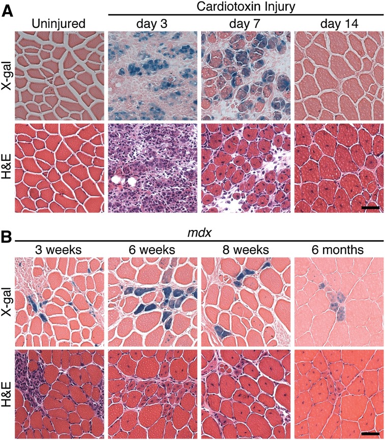
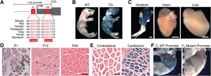
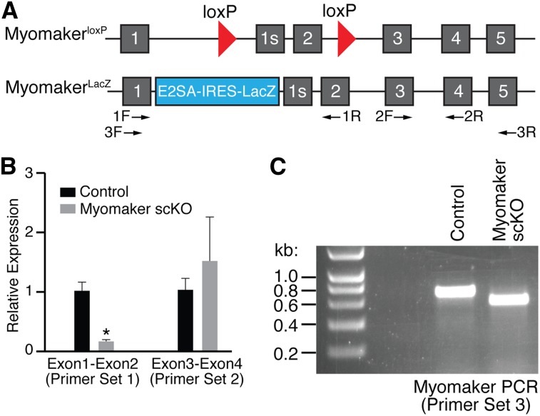
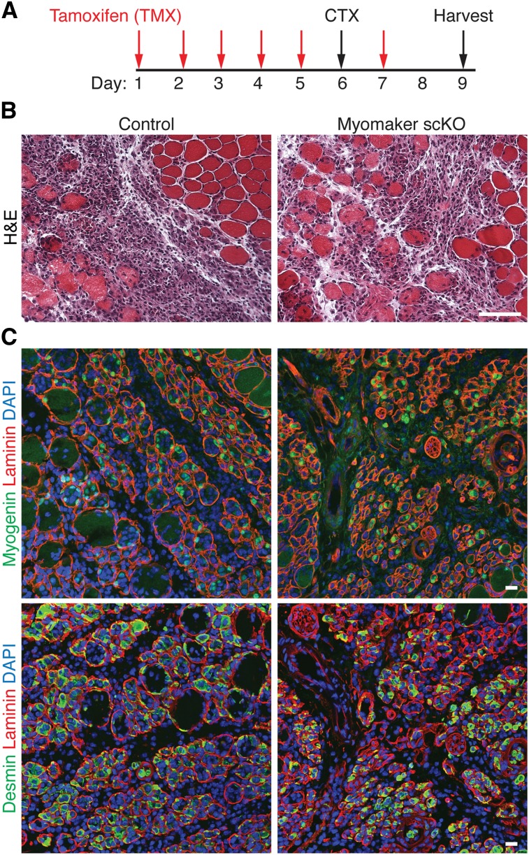
Regeneration of injured adult skeletal muscle involves fusion of activated satellite cells to form new myofibers. Myomaker is a muscle-specific membrane protein required for fusion of embryonic myoblasts, but its potential involvement in adult muscle regeneration has not been explored. We show that myogenic basic helix-loop-helix (bHLH) transcription factors induce myomaker expression in satellite cells during acute and chronic muscle regeneration. Moreover, genetic deletion of myomaker in adult satellite cells completely abolishes muscle regeneration, resulting in severe muscle destruction after injury. Myomaker is the only muscle-specific protein known to be absolutely essential for fusion of embryonic and adult myoblasts.

Keywords: E-box elements; fusion; muscle regeneration; myogenesis; myomaker.


Figures

Figure 1.
Figure 1.
Transient expression of myomaker during acute and chronic muscle injury. (A) The TA muscle from myomakerLacZ/+ mice was injured with CTX, cryosectioned, and stained with X-gal. Myomaker-lacZ is expressed at days 3 and 7 post-injury and then down-regulated. (B) MyomakerLacZ/+ mice were crossed with mdx mice. TA muscles at the indicated time points were analyzed for myomaker expression through X-gal staining. For A and B, serial hematoxylin and eosin (H&E) sections accompany each X-gal image. Bar, 50 μm.
Figure 2.
Figure 2.
Myomaker expression is controlled by promoter E-box elements. (A) Schematic of 1.7-kb myomaker promoter with proximal E-boxes notated. Mammalian conservation is shown below the schematic. The 1.7-kb promoter was cloned upstream of LacZ, and stable transgenic mice were generated. (B) Postnatal day 3 (P3) wild-type (WT) and transgenic mice stained with X-gal. Bar, 1 mm. (C) β-Galactosidase activity was detected in P3 hindlimbs but absent in heart and liver. Bar, 1 mm. (D) The myomaker promoter is expressed in muscle fibers at P1, is expressed to a lesser extent at P14, and is inactive at P28. Bar, 50 μm. (E) Transgenic mice harboring the myomaker promoter were subjected to CTX and stained with X-gal; the promoter is activated during regeneration. Bar, 50 μm. (F) The E-boxes (+32 base pairs [bp] and −41 bp) were mutated within the 1.7-kb promoter and assayed for the ability to drive LacZ expression in F0 transgenic mice. The wild-type promoter showed LacZ expression in eight of 10 P1 mice, but the mutant promoter lacked activity. Bar, 1 mm.
Figure 3.
Figure 3.
Analysis of myomaker transcript after tamoxifen-induced satellite cell deletion. (A) Diagram showing the myomaker targeted alleles used in this study. The myomakerloxP allele contains loxP sites flanking exon 2, and the myomakerLacZ/+ allele has a LacZ cassette in intron 1 that inhibits exon 1 splicing with exon 2. Exon 1s refers to the first exon of a short isoform of myomaker that is annotated but not highly conserved within mammalian species. The arrows below the schematic denote primer sets used in B and C. (B) Both control (n = 3, myomakerLacZ/+;Pax7-CreERT2 or myomakerloxP/+;Pax7-CreERT2) and myomakerscKO (n = 4, myomakerLacZ/loxP;Pax7-CreERT2) mice were treated with tamoxifen, injured with CTX, and analyzed by qPCR for the presence of exons 1 and 2 and exons 3 and 4 using the indicated primer sets. Data are presented as mean ± SEM. (*) P < 0.05. (C) PCR with primer set 3 shows a shorter amplicon in myomakerscKO muscle due to the Cre-mediated excision of exon 2.
Figure 4.
Figure 4.
Loss of myomaker in adult satellite cells does not alter the early response to muscle injury. (A) Schematic showing the timing of tamoxifen (TMX) and CTX. Injured muscle was analyzed 3 d after CTX injection. (B) Representative H&E-stained sections from control and myomakerscKO muscle demonstrate similar levels of damage. Bar, 50 μm. (C) Myogenin and desmin immunohistochemistry indicates the presence of myogenic cells in myomakerscKO muscle. Bar, 20 μm.
Figure 5.
Figure 5.
Myomaker is necessary for muscle regeneration. (A) Time course of tamoxifen (TMX) and CTX treatment. TA muscles were analyzed 9 d post-injury. (B) H&E-stained sections from control and myomakerscKO mice show a complete lack of regeneration after genetic deletion of myomaker in satellite cells. Bars: top image, 500 μm; bottom image, 100 μm. (C) Myosin and desmin staining revealed a dramatic loss of muscle cells and no regenerated muscle fibers in myomakerscKO TA. Bar, 20 μm.

References

    1. Berkes CA, Tapscott SJ. 2005. MyoD and the transcriptional control of myogenesis. Semin Cell Dev Biol 16: 585–595 - PubMed
    1. Buckingham M 2006. Myogenic progenitor cells and skeletal myogenesis in vertebrates. Curr Opin Genet Dev 16: 525–532 - PubMed
    1. Charrin S, Latil M, Soave S, Polesskaya A, Chretien F, Boucheix C, Rubinstein E. 2013. Normal muscle regeneration requires tight control of muscle cell fusion by tetraspanins CD9 and CD81. Nat Commun 4: 1674. - PubMed
    1. Doherty KR, Cave A, Davis DB, Delmonte AJ, Posey A, Earley JU, Hadhazy M, McNally EM. 2005. Normal myoblast fusion requires myoferlin. Development 132: 5565–5575 - PMC - PubMed
    1. Durbeej M, Campbell KP. 2002. Muscular dystrophies involving the dystrophin-glycoprotein complex: an overview of current mouse models. Curr Opin Genet Dev 12: 349–361 - PubMed

Publication types