. 2014 Aug 1:7:54.
doi: 10.1186/s13041-014-0054-1.

The Amyloid Precursor Protein is rapidly transported from the Golgi apparatus to the lysosome and where it is processed into beta-amyloid

Affiliations

The Amyloid Precursor Protein is rapidly transported from the Golgi apparatus to the lysosome and where it is processed into beta-amyloid

Joshua H K Tam et al. Mol Brain. .

Abstract

Background: Alzheimer's disease (AD) is characterized by cerebral deposition of β-amyloid peptide (Aβ). Aβ is produced by sequential cleavage of the Amyloid Precursor Protein (APP) by β- and γ-secretases. Many studies have demonstrated that the internalization of APP from the cell surface can regulate Aβ production, although the exact organelle in which Aβ is produced remains contentious. A number of recent studies suggest that intracellular trafficking also plays a role in regulating Aβ production, but these pathways are relatively under-studied. The goal of this study was to elucidate the intracellular trafficking of APP, and to examine the site of intracellular APP processing.

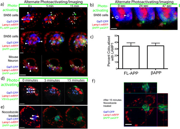
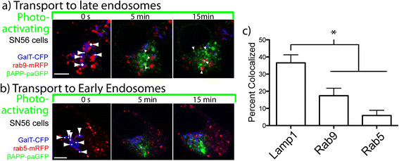
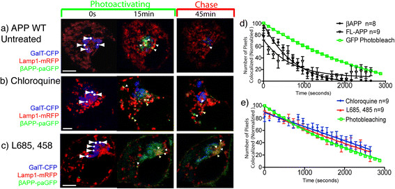
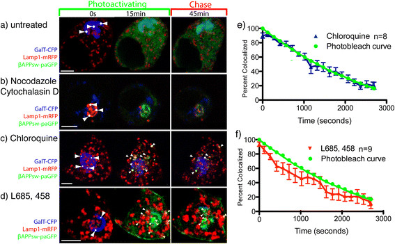
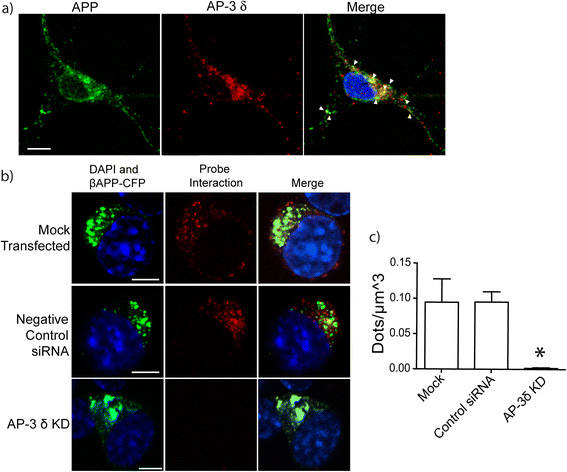
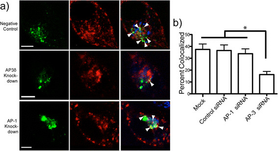
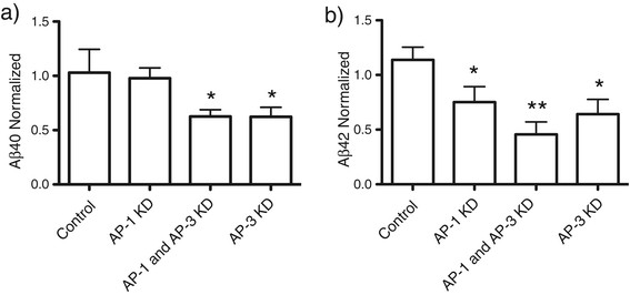
Results: We have tagged APP on its C-terminal cytoplasmic tail with photoactivatable Green Fluorescent Protein (paGFP). By photoactivating APP-paGFP in the Golgi, using the Golgi marker Galactosyltranferase fused to Cyan Fluorescent Protein (GalT-CFP) as a target, we are able to follow a population of nascent APP molecules from the Golgi to downstream compartments identified with compartment markers tagged with red fluorescent protein (mRFP or mCherry); including rab5 (early endosomes) rab9 (late endosomes) and LAMP1 (lysosomes). Because γ-cleavage of APP releases the cytoplasmic tail of APP including the photoactivated GFP, resulting in loss of fluorescence, we are able to visualize the cleavage of APP in these compartments. Using APP-paGFP, we show that APP is rapidly trafficked from the Golgi apparatus to the lysosome; where it is rapidly cleared. Chloroquine and the highly selective γ-secretase inhibitor, L685, 458, cause the accumulation of APP in lysosomes implying that APP is being cleaved by secretases in the lysosome. The Swedish mutation dramatically increases the rate of lysosomal APP processing, which is also inhibited by chloroquine and L685, 458. By knocking down adaptor protein 3 (AP-3; a heterotetrameric protein complex required for trafficking many proteins to the lysosome) using siRNA, we are able to reduce this lysosomal transport. Blocking lysosomal transport of APP reduces Aβ production by more than a third.

Conclusion: These data suggests that AP-3 mediates rapid delivery of APP to lysosomes, and that the lysosome is a likely site of Aβ production.


Figures

Figure 1
Figure 1
Overview of constructs. For these experiments, APP constructs were generated including the full length APP 751 fused to paGFP on its C-terminus. A shorter construct consisting of the C-terminal 112 amino acids of APP fused to paGFP. Both constructs include a linker with includes an N-terminal HA epitope tag, and both constructs contain α-, β- and γ- cleavage sites. Cleavage at the γ-site will release the C-terminal tail of APP along with the paGFP tag into the cytoplasm.
Figure 2
Figure 2
APP is rapidly trafficked from the Golgi apparatus to LAMP1-labeled compartment. Photoactivation targets were drawn on the Golgi apparatus (white dots with arrows). For 15 minutes, cells were alternately imaged and irradiated (photoactivated) with 405 nm laser light within the targets. White arrowheads point to APP-paGFP colocalized with Lamp1-mRFP. Scale Bar = 5 μm. a) Demonstrates rapid transport of Full length-APP- paGFP (top panel) and βAPP-paGFP (middle panel) from Golgi apparatus to a LAMP1-labeled compartment. The same trafficking occurs in mouse primary neurons (lower panel). (See Additional file 3: Video S1). b) Higher magnification images of βAPP-paGFP trafficking rapidly to a LAMP1 labeled compartment. Scale Bar = 1 μm. c) Comparison of the colocalization of activated FL-APP-paGFP (n = 9) and activated βAPP-paGFP (n = 8). d) SN56 cells transiently transfected with the secretory protein Vesicular Stomatitis Virus Glycoprotein-paGFP (VSVG-paGFP), GalT-CFP (blue), and LAMP1-mRFP (red) to demonstrate that very little of the green photoactivated VSVG-paGFP arrives in the LAMP1 compartment; paGFP does not alter trafficking. Scale bars = 5 μm. e) Transfected SN56 cells were treated for 5 minutes with nocodazole before imaging (See Additional file 5: Video S2) Scale bars = 5 μm. f) Z-stack of the same cell taken immediately following 15 minutes of photoactivation demonstrating that green signal remains inside the Golgi.
Figure 3
Figure 3
APP is primarily transported to a LAMP1 compartment. SN56 cells were cotransfected with plasmids expressing APP-paGFP, GalT-CFP (blue), and a compartment marker (red). Photoactivation targets were drawn on the Golgi apparatus (white dots with arrows). βAPP-paGFP trafficking was visualized from the from Golgi apparatus to Rab 9 labelled late endosomes (a) and Rab 5 labelled early endosomes (b). Scale bars represent 5 μm. c) Percent of APP-paGFP fluorescence colocalized with respective compartment markers after 15 minutes of photoactivation in the Golgi (circles: LAMP1 (n = 9), squares: Rab9 (n = 10), triangles: Rab5 (n = 7)). Error bars represent standard deviation. (* = p < 0.05).
Figure 4
Figure 4
APP is processed in the lysosome by a γ-secretase like activity. SN56 cells were transiently transfected with βAPP-paGFP, GalT-CFP, and LAMP1-mRFP. Cells were alternately photoactivated with 405 nm light and imaged in the Golgi for 15 minutes, and then imaged every 30 seconds for 1 hour. a) Shows the accumulation of photoactivated APP-paGFP in the lysosome after 15 minutes, follow by its near complete clearance after 45 minutes. Arrowheads denote areas of colocalization. (See also Additional file 3: Video S1) b) Transiently transfected SN56 cells were pretreated with 100 μM chloroquine for 30 minutes prior to imaging. After chloroquine treatment APP is still visible in lysosomes after 45 minutes (See Additional file 6: Video S3). c) Cells treated with 0.5 μM L685, 458 (γ-secretase inhibitor) overnight prior to photoactivating/imaging. L685, 458 treatment substantially increases the accumulation of photoactivated βAPP-paGFP in lysosomes, and substantially decreases its cleance. Scale bars represent 5 μm (See Additional file 7: Video S4). d) Cleavage of βAPP-paGFP was determined by measuring the loss of FL-APP (black open triangles) and βAPP-paGFP (black closed circles) from LAMP1 labeled compartments. Values were averaged and normalized to begin at 100%. Overlaid in green squares is the loss of fluorescence of EGFP in the identical imaging protocol. Error bars represent SEM. (* = p < 0.05) (e) Shows the clearance of photoactivated APP-paGFP cells that were treated with 100 μM chloroquine for 30 minutes before imaging (n = 9) or with 0.5 μM L685, 458 (γ-secretase inhibior) (n = 9). Error bars represent SEM.
Figure 5
Figure 5
The Swedish mutation causes rapid clearance of APP from lysosomes. SN56 cells were transiently transfected with βAPPsw-paGFP, GalT-CFP, and LAMP1-mRFP. Scale bars represent 5 μm. a) βAPPsw-paGFP was photoactivated for 15 minutes in the GalT-CFP labeled compartment, and chased for 45 minutes. βAPPsw is cleaved nearly instantaneously and appears in the cytoplasm. b) Cells were treated for 5 minutes before imaging with 66 μM nocodazole and 10 μM cytochalasin. GalT-CFP is false colored red to provide better contrast, and LAMP1-mRFP has been false coloured blue. Photoactivated βAPPsw-paGFP accumulates in the Golgi and does not appear to be cleaved. c) Cells were treated acutely with 100 μM chloroquine (See Additional file 10: Video S7) which results in photoactivated βAPPsw-paGFP accumulating in lysosomes. White arrowheads represent βAPPsw-paGFP colocalized with LAMP1-mRFP d) Cells were treated with 0.5 μM L658, 458 (See Additional file 11: Video S8), which also causes photoactivated βAPPsw-paGFP to appear in lysosomes. Scale bars represent 5 μm. Quantitation of colocalized green pixels with LAMP1-mRFP show that the clearance of βAPPsw-paGFP from the lysosome proceeds linearly after treatment with e) chloroquine (n = 8), or with f) L658, 458 (n = 9). Error bars represent standard deviation.
Figure 6
Figure 6
AP-3δ and APP colocalize and interact. a) E15 mouse neurons were cultured and immunostained with antibodies against AP-3δ (SA4; red) and APP (APP C-terminal; green). Arrowheads point to colocalized pixels. Scale bars represent 5 μm. Inset shows magnified view of the cell body. b) Proximity ligation assay (PLA) demonstrates the interaction of APP and AP-3δ. Cells were transiently transfected with βAPP-CFP with no siRNA, control siRNA or AP-3δ siRNA. Cells were stained with mouse anti- AP-3δ and rabbit anti-APP C-terminal antibodies. These were detected with secondary antibodies conjugated to complementary DNA sequences. When proteins are within 40 nm, DNA is ligated and replicated and detected by in-situ fluorescent red dots. AP-3δ siRNA substantially reduces the number of red dots. (scale bars represent 10 μm) c) Quantification of PLA fluorescent dots in SN56 cells normalized to cell volume (*p < 0.05).
Figure 7
Figure 7
AP-3 mediates direct trafficking of APP to lysosomes. a) SN56 cells were transfected with βAPP-paGFP, LAMP1-mRFP, GalT-CFP, and either control siRNA, siRNA against AP-3δ mRNA or siRNA against AP-1γ. Cells were alternately photoactivated with 405 nm light and imaged in the Golgi for 15 minutes (scale bar represents 5 μm). White arrowheads in the merged image (far right panel) denote colocalized pixels. Scale bars represent 5 μm. b) Percent of βAPP-paGFP colocalizing with LAMP1-mRFP at the end of the 15-minute photoactivation period. (* = p < 0.05; Error bars represent standard deviation).
Figure 8
Figure 8
AP-3 mediates processing to Aβ. SN56 cells were transfected with βAPP-paGFP, LAMP1-mRFP, GalT-CFP, and either control siRNA, siRNA against AP-3δ mRNA or siRNA against AP-1γ. a) SN56 cells were co-transfected a plasmid expressing βAPPswe-CFP and with control siRNAs, siRNA against AP-1γ, siRNA against AP-1γ and AP-3δ combined, or siRNA against AP-3δ. Conditioned media was analyzed for a) Aβ40 or b) Aβ42 by ELISA. Experiments were performed 4 times, with each experiment consisting of 2 replicates. (* indicates significantly different from control p < 0.05; ** indicates significantly different from control and either AP-1 or AP-3 alone p < 0.05) Error bars represent SEM.

Similar articles

Cited by

References

    1. Mebane-Sims I. Alzheimer's disease facts and figures. Alzheimers Dement. 2009;2009(5):234–270. - PubMed
    1. Nelson PT, Braak H, Markesbery WR. Neuropathology and cognitive impairment in Alzheimer disease: a complex but coherent relationship. J Neuropathol Exp Neurol. 2009;68:1–14. - PMC - PubMed
    1. Lin X, Koelsch G, Wu S, Downs D, Dashti A, Tang J. Human aspartic protease memapsin 2 cleaves the beta-secretase site of beta-amyloid precursor protein. Proc Natl Acad Sci USA. 2000;97:1456–1460. - PMC - PubMed
    1. Hussain I, Powell D, Howlett DR, Tew DG, Meek TD, Chapman C, Gloger IS, Murphy KE, Southan CD, Ryan DM, Smith TS, Simmons DL, Walsh FS, Dingwall C, Christie G. Identification of a Novel Aspartic Protease (Asp 2) as β-Secretase. Mol Cell Neurosci. 1999;14:419–427. - PubMed
    1. Vassar R, Bennett BD, Babu-Khan S, Kahn S, Mendiaz EA, Denis P, Teplow DB, Ross S, Amarante P, Loeloff R, Luo Y, Fisher S, Fuller J, Edenson S, Lile J, Jarosinski MA, Biere AL, Curran E, Burgess T, Louis JC, Collins F, Treanor J, Rogers G, Citron M. Beta-secretase cleavage of Alzheimer's amyloid precursor protein by the transmembrane aspartic protease BACE. Science. 1999;286:735–741. - PubMed

Publication types

Substances

LinkOut - more resources