Review
. 2014 Aug 1;6(8):a016972.
doi: 10.1101/cshperspect.a016972.

Endocytosis of viruses and bacteria

Affiliations
Review

Endocytosis of viruses and bacteria

Pascale Cossart et al. Cold Spring Harb Perspect Biol. .

Abstract

Of the many pathogens that infect humans and animals, a large number use cells of the host organism as protected sites for replication. To reach the relevant intracellular compartments, they take advantage of the endocytosis machinery and exploit the network of endocytic organelles for penetration into the cytosol or as sites of replication. In this review, we discuss the endocytic entry processes used by viruses and bacteria and compare the strategies used by these dissimilar classes of pathogens.


Figures

Figure 1.
Figure 1.
Viruses in the endocytic network. After binding to cell-surface receptors, viruses are internalized through a variety of endocytic processes including macropinocytosis, clathrin-mediated endocytosis, caveolae, and clathrin- and caveolin-independent mechanisms. Some of the viruses that use these mechanisms of endocytosis are listed. The primary endocytic vesicles and vacuoles formed ferry incoming virus particles into the endocytic network, where they undergo sorting and eventually penetration into the cytosol from different locations within the vacuolar network. Five different locations are indicated by the numbered gray arrows. Pathways followed by influenza A virus, SV40, Uukuniemi virus, and vaccinia virus are shown. Of these, SV40 is transported to the endoplasmic reticulum for uncoating and penetration, whereas influenza A and vaccinia virus undergo acid-activated membrane fusion in maturing and late endosomes and macropinosomes, respectively. The location and timing of escape are often determined by the pH threshold for penetration of the virus particles as indicated. LCMV, lymphocytic choriomeningitis virus.
Figure 2.
Figure 2.
Pathways of endocytic entry by invasive bacteria. Schematic representation of bacterial endocytosis mechanisms and intravacuolar or intracytosolic lifestyles. In epithelial cells, Listeria enters via two internalins; it then resides transiently in a vacuole that is lysed allowing intracytosolic replication and actin-based motility. The obligate intracellular bacterium Chlamydia trachomatis resides in a vacuole that intercepts the pathway involved in the transport of shingomyelin from the Golgi apparatus to the plasma membrane. Shigella is first taken up by filopodia present on the surface of epithelial cells. The type 3SS then injects effectors that trigger entry, formation of the vacuole, and escape from this compartment, leading to intracytosolic replication and actin-based motility. Salmonella appears to enter after a “near-surface swimming” mechanism. The T3SS-1 (or SPI-1) triggers entry and formation of the replicative vacuole, which acquires markers of endosomes and lysosomes owing to the secretion of effectors of the T3SS-2 (or SPI-2). In some circumstances, Salmonella can reach the cytosol and rapidly replicate therein. The Brucella replicative vacuole is derived from the endocytic vacuole and matures in an ER-derived vacuole. Coxiella burnetii is the only bacterium that has evolved to survive and replicate in a lysosome-derived vacuole. Bartonella henselae can enter into endothelial cells as a single bacterium or as a group, leading to the formation of an invasome. In macrophages, Legionella and Mycobacterium tuberculosis reside in a vacuole. The Legionella vacuole acquires markers of the ER. It is not the case for M. tuberculosis, which blocks the maturation of the internalization vacuole.
Figure 3.
Figure 3.
Semliki Forest virus is internalized by clathrin-coated vesicles. After attaching to the surface of BHK-21 cells, this simple, enveloped RNA virus of the toga(α)virus family is rapidly endocytosed by clathrin-mediated endocytosis, and penetration occurs in early endosomes (Helenius et al. 1980). Scale bar, 200 nm.
Figure 4.
Figure 4.
Endocytosis of enveloped and nonenveloped viruses. These electron micrographs show viruses from different families during endocytic entry. (A) Bound to its ganglioside receptors (GD1a and GT1b), an incoming mouse polyoma virus particle is seen in a tight-fitting indentation of the plasma membrane of a 3T6 cell. Although viruses of the polyoma virus family can also use caveolae (see arrow) for entry, this particle is most likely making use of a clathrin- and caveolin-independent mechanism used by these viruses. (B) Human papilloma virus 16 enters HeLa cells via a macropinocytosis-like mechanism that involves binding to filopodia, surfing along filopodia to the cell body, and activation of an endocytic process independent of clathrin and caveolin (Schelhaas et al. 2008, 2012). (C) Vesicular stomatitis virus is internalized by clathrin-coated vesicles (Matlin et al. 1982). (D) An influenza A virus particle is seen in an early endosome after endocytic internalization (Matlin et al. 1981). (E) In this cryoEM image, a vaccinia virus particle (a poxvirus) is seen in a macropinocytic vacuole after endocytosis in a HeLa cell. The particle is labeled with immunogold against the A5 core protein. (F) The contact between an SV40 particle and the plasma membrane is very tight during the clathrin- and caveolin-independent entry process. The virus seems to bud into the cell. Scale bars, 200 nm. The images are by Roberta Mancini (A–D) and Christopher Bleck (E).
Figure 5.
Figure 5.
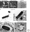
Endocytosis of bacteria. These electron micrographs show various phases of bacterial endocytosis into mammalian cells. (A, left) Binding of Listeria entering into cells. Two coated pits are detectable on this cross section. The tight apposition of the membrane illustrates what is meant by the zipper mechanism. (A, middle micrograph) A Shigella entering into a cell and the huge membrane ruffles that engulf the bacterium. This micrograph illustrates the trigger mechanism. (A, right) Bartonella henselae enter as a group into an endothelial cell. (B) Listeria is entering into the cell and the clathrin coat is visible as a thickening of the plasma membrane underneath the bacterium. (C, left) Listeria is present in a membrane-bound vacuole. (C, right) Chlamydia vacuole full of bacteria that have replicated.

References

    1. Agbor TA, McCormick BA 2011. Salmonella effectors: Important players modulating host cell function during infection. Cell Microbiol 13: 1858–1869 - PMC - PubMed
    1. Anderson HA, Chen Y, Norkin LC 1996. Bound simian virus 40 translocates to caveolin-enriched membrane domains, and its entry is inhibited by drugs that selectively disrupt caveolae. Mol Biol Cell 7: 1825–1834 - PMC - PubMed
    1. Backert S, Meyer TF 2006. Type IV secretion systems and their effectors in bacterial pathogenesis. Curr Opin Microbiol 9: 207–217 - PubMed
    1. Bakowski MA, Braun V, Lam GY, Yeung T, Heo WD, Meyer T, Finlay BB, Grinstein S, Brumell JH 2010. The phosphoinositide phosphatase SopB manipulates membrane surface charge and trafficking of the Salomnella-containg vacuole. Cell Host Microbe 7: 453–462 - PubMed
    1. Bastidas RJ, Elwell CA, Engel JN, Valdivia RH 2013. Chlamydial intracellular survival strategies. Cold Spring Harb Perspect Med 3: a010256. - PMC - PubMed

Publication types