

Mucoepidermoid carcinoma
- PMID: 25085946
- PMCID: PMC4127757
- DOI: 10.1136/bcr-2013-202776
Mucoepidermoid carcinoma
Abstract
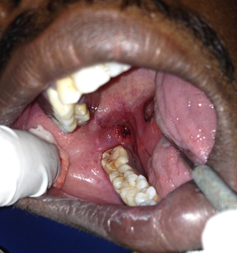
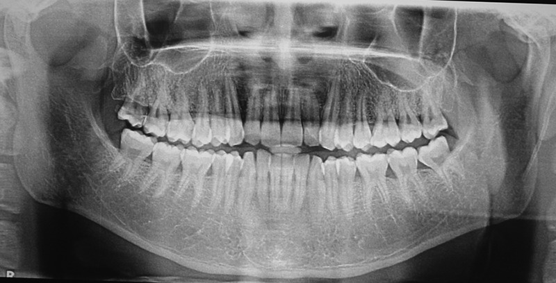
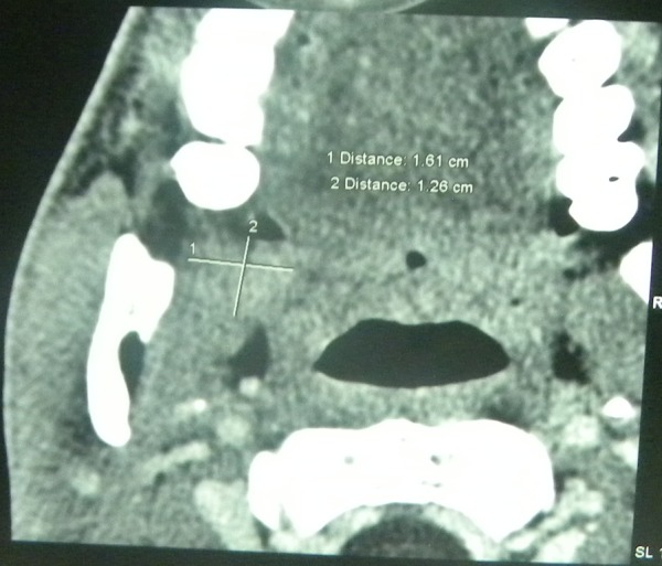
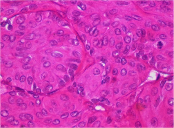
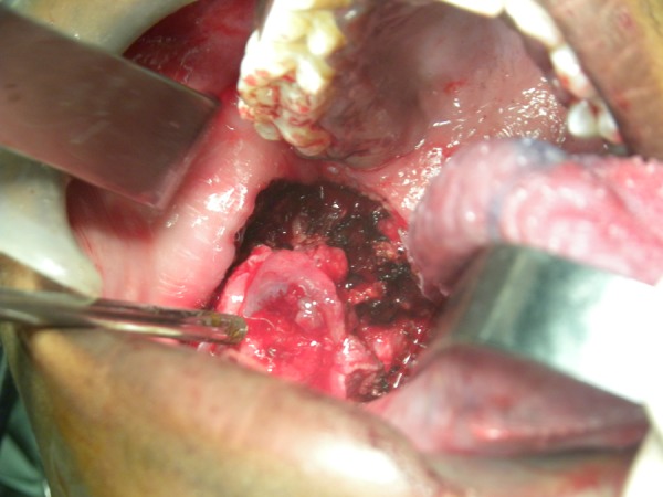
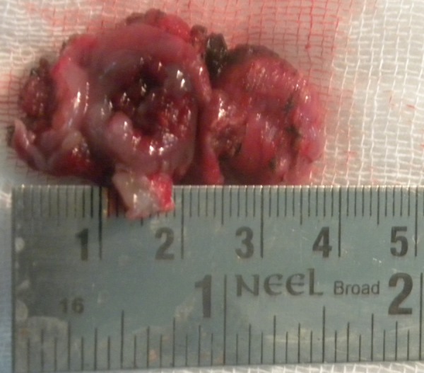
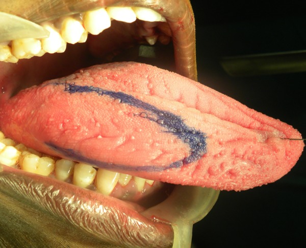
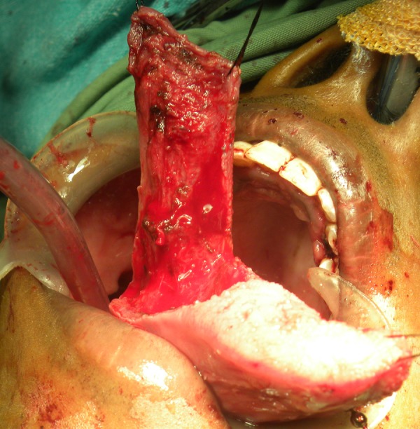
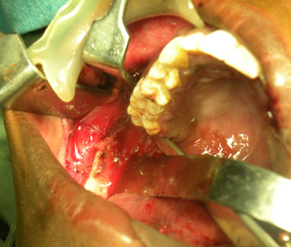
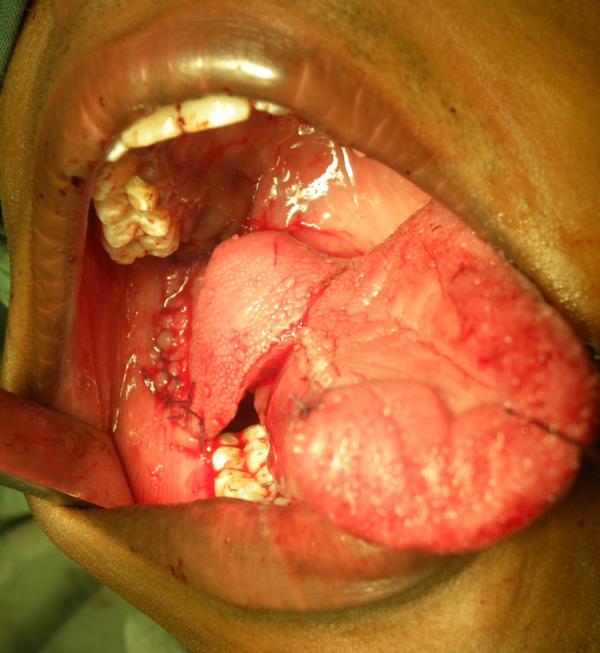
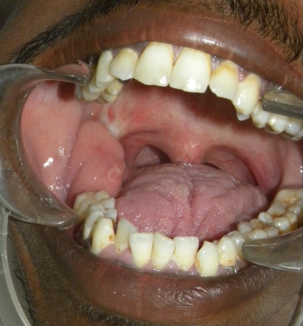
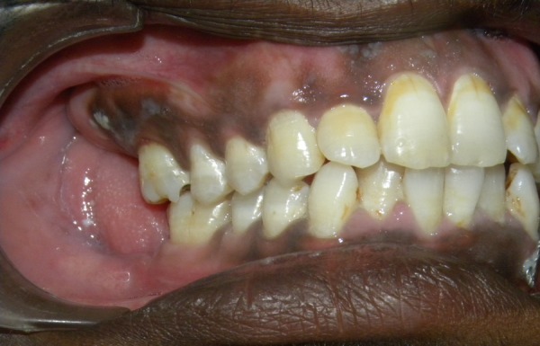
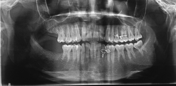
Salivary gland tumours comprise almost 5% of head and neck malignancies. Minor salivary gland tumours account for 10-15% of all salivary gland neoplasms and are usually malignant. The second most common minor salivary gland tumour (12-40% globally) is mucoepidermoid carcinoma. Mucoepidermoid carcinoma is more frequent in females, occurs in the fifth decade of life and is usually found in the parotid gland. However, the palate is a frequent site when it occurs in the minor glands. We report a case of a high-grade variant of mucoepidermoid carcinoma in the right retromolar trigone of a 21-year man which was treated with wide excision of the tumour with a 1.5 cm margin. Reconstruction was done with a buccal fat pad posteriorly with a pedicled lateral tongue flap. Temporal stripping and right coronoidectomy was carried out in case of post-surgical wound contraction. The patient is currently under periodic review.
2014 BMJ Publishing Group Ltd.
Figures
References
-
- Lutcavage GJ, Schaberg SJ, Fulbright DK, et al. Retromolar trigone mass. J Oral Maxillofac Surg 1993;51:1024–9 - PubMed
-
- Wood NK, Goaz PW, Kallal R. Multilocular Radiolucencies in Differential Diagnosis of Oral and Maxillofacial Lesions 5th edn 2006:346–7
-
- Pires FR, Chen SY, Da Cruz Perez DE, et al. Cytokeratin expression in central mucoepidermoid carcinoma and glandular odontogenic cyst. Oral Oncology 2004;40:545–51 - PubMed
Publication types
MeSH terms
LinkOut - more resources
Full Text Sources
Other Literature Sources
Medical