. 2014 Sep 19;289(38):26021-26037.
doi: 10.1074/jbc.M114.561134. Epub 2014 Aug 1.

Nrbf2 protein suppresses autophagy by modulating Atg14L protein-containing Beclin 1-Vps34 complex architecture and reducing intracellular phosphatidylinositol-3 phosphate levels

Affiliations

Nrbf2 protein suppresses autophagy by modulating Atg14L protein-containing Beclin 1-Vps34 complex architecture and reducing intracellular phosphatidylinositol-3 phosphate levels

Yu Zhong et al. J Biol Chem. .

Abstract

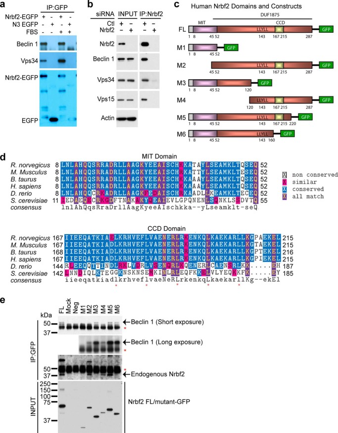
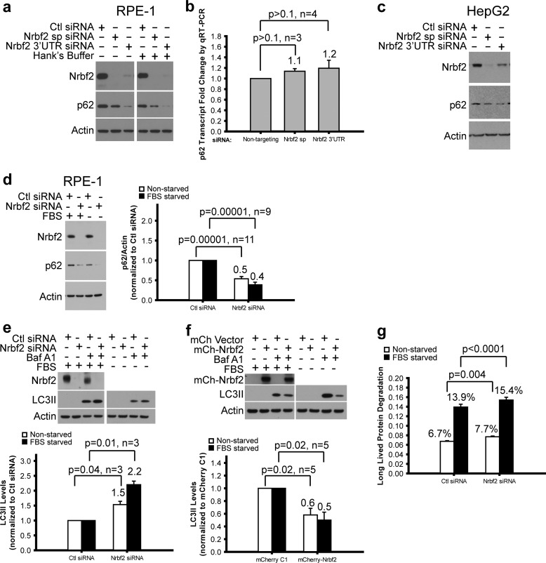
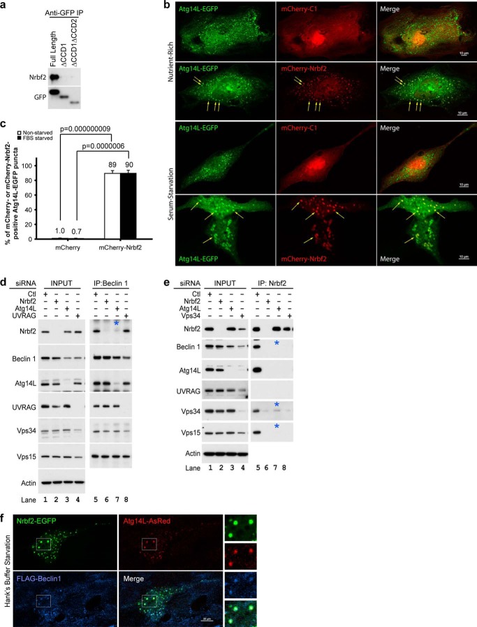
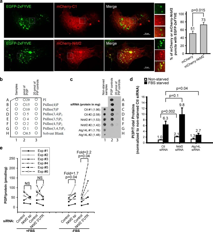
Autophagy is a tightly regulated lysosomal degradation pathway for maintaining cellular homeostasis and responding to stresses. Beclin 1 and its interacting proteins, including the class III phosphatidylinositol-3 kinase Vps34, play crucial roles in autophagy regulation in mammals. We identified nuclear receptor binding factor 2 (Nrbf2) as a Beclin 1-interacting protein from Becn1(-/-);Becn1-EGFP/+ mouse liver and brain. We also found that Nrbf2-Beclin 1 interaction required the N terminus of Nrbf2. We next used the human retinal pigment epithelial cell line RPE-1 as a model system and showed that transiently knocking down Nrbf2 by siRNA increased autophagic flux under both nutrient-rich and starvation conditions. To investigate the mechanism by which Nrbf2 regulates autophagy, we demonstrated that Nrbf2 interacted and colocalized with Atg14L, suggesting that Nrbf2 is a component of the Atg14L-containing Beclin 1-Vps34 complex. Moreover, ectopically expressed Nrbf2 formed cytosolic puncta that were positive for isolation membrane markers. These results suggest that Nrbf2 is involved in autophagosome biogenesis. Furthermore, we showed that Nrbf2 deficiency led to increased intracellular phosphatidylinositol-3 phosphate levels and diminished Atg14L-Vps34/Vps15 interactions, suggesting that Nrbf2-mediated Atg14L-Vps34/Vps15 interactions likely inhibit Vps34 activity. Therefore, we propose that Nrbf2 may interact with the Atg14L-containing Beclin 1-Vps34 protein complex to modulate protein-protein interactions within the complex, leading to suppression of Vps34 activity, autophagosome biogenesis, and autophagic flux. This work reveals a novel aspect of the intricate mechanism for the Beclin 1-Vps34 protein-protein interaction network to achieve precise control of autophagy.

Keywords: Autophagosome Biogenesis; Autophagy; Beclin 1; Cellular Regulation; Nrbf2; Protein Degradation; Protein-Protein Interaction; Subcellular Organelle; Vps34.


Figures

FIGURE 1.
FIGURE 1.
Identification of Nrbf2 as a novel Beclin 1-interacting protein. a, anti-EGFP antibody pulled down endogenous Beclin 1 and Vps34 in HEK293 cells stably expressing mouse Nrbf2-EGFP under both nutrient-rich and serum starvation conditions. Immunoprecipitated Nrbf2-EGFP and EGFP were labeled in the anti-GFP blot. b, anti-Nrbf2 antibody pulled down endogenous Beclin 1, Vps34, and Vps15 from RPE-1 cells transfected with non-targeting siRNA but not from those transfected with the SMARTpool Nrbf2 siRNA. Of note, both cell extracts before IP (INPUT) contain similar levels of Beclin 1, Vps34, Vps15, and actin. c, human Nrbf2 domain structures and diagrams of the C-terminal Cycle3 GFP-tagged full-length (FL) human Nrbf2 and truncation mutants (M1-M6) of Nrbf2. d, sequence alignment of the Nrbf2 MIT and CCD domains from different species. The five conserved leucines in the CCD are marked by red asterisks. Note that Atg38, the yeast ortholog of Nrbf2, has low sequence homology to the mammalian Nrbf2, particularly lacking the abovementioned five leucines in the CCD. e, anti-GFP IP of Cycle3 GFP-tagged full-length and mutant human Nrbf2 constructs from HepG2 cells revealed that the N-terminal 120 residues of Nrbf2 were required and sufficient for the Nrbf2-Beclin 1 interaction. Red asterisks mark the IgG bands. GFP-tagged full-length and mutant Nrbf2 constructs in the INPUT were probed with anti-GFP antibody.
FIGURE 2.
FIGURE 2.
Characterization of Nrbf2 as a negative regulator of autophagic flux under both nutrient-rich and starvation conditions in human cell lines. Western blot analysis of p62 in RPE-1 (a) and HepG2 (c) cells transfected with non-targeting control (Ctl) siRNA, human SMARTpool (sp) and 3′UTR Nrbf2 siRNAs showed decreased p62 levels upon Nrbf2 siRNA treatment. a, cells were grown under nutrient-rich or 1-h Hanks' buffer starvation conditions. c, cells were grown under nutrient-rich conditions. b, qRT-PCR quantification of the relative p62 transcript levels in the RPE-1 cells transfected with Ctl, Nrbf2 sp, and Nrbf2 3′UTR siRNAs. d, Western blot analysis of p62 levels in RPE-1 cells transfected with Ctl or Nrbf2 sp siRNA showed decreased p62 levels upon Nrbf2 siRNA treatment under both nutrient-rich and 18-h serum starvation conditions. e, Western blot analysis of LC3II levels in RPE-1 cells transfected with Ctl siRNA or Nrbf2 sp siRNA and treated either without or with 200 nm Baf showed a greater Baf-induced increase of LC3II levels upon Nrbf2 siRNA treatment under both nutrient-rich and 18-h serum starvation conditions. f, Western blot analysis of LC3II levels in RPE-1 cells transfected with mCherry empty vector or mCherry-tagged human Nrbf2 and treated either without or with 200 nm Baf showed a lesser Baf-induced increase of LC3II levels upon Nrbf2 overexpression under both nutrient-rich and 18-h serum starvation conditions. The statistical significance for the LC3II assays in e and f is lower than that for the p62 assay in d, likely because of fewer numbers of independent experiments. g, the long-lived protein degradation rate increased upon Nrbf2 siRNA treatment under both nutrient-rich and serum starvation conditions. For all quantifications, means ± S.E. were plotted with means labeled.
FIGURE 3.
FIGURE 3.
Nrbf2 is a component of the Atg14L-containing Beclin 1-Vps34 protein complex. a, transiently expressed, EGFP-tagged, full-length Atg14L, but not the CCD-deletion mutants of Atg14L, pulled down endogenous Nrbf2 in HeLa cells. Immunoprecipitated EGFP-tagged full-length and mutant Atg14L were probed with anti-GFP antibody. b, ectopically expressed mCherry-tagged human Nrbf2 colocalized with Atg14L-EGFP on punctate structures (arrows) in RPE-1 cells under nutrient-rich and 18-h serum-starvation conditions. In contrast, mCherry alone showed diffused cytoplasmic and nuclear localization. Scale bars = 10 μm. c, quantification of colocalization of mCherry or mCherry-Nrbf2 with Atg14L-EGFP as measured by the percentage of Atg14L-EGFP puncta that were either mCherry- or mCherry-Nrbf2-positive. Cells with more than five Atg14L-EGFP puncta were used for quantification. The numbers of cells used for quantification were five (for mCherry, nutrient-rich), seven (for mCherry, serum starvation), nine (for mCherry-Nrbf2, nutrient-rich), and seven (for mCherry-Nrbf2, serum starvation). These cell numbers are sufficient to reveal a statistical significance of colocalization between mCherry-Nrbf2 and Atg14L-EGFP using two-tailed Student's t test. d, anti-Beclin 1 IP from RPE-1 cell treated with non-targeting, Nrbf2, Atg14L, or UVRAG siRNA. e, anti-Nrbf2 IP from RPE-1 cells treated with non-targeting, Nrbf2, Atg14L, or Vps34 siRNA. Note that Vps34 siRNA treatment led to unstable Nrbf2, Beclin 1, Atg14L, UVRAG, and Vps15, whereas Atg14L siRNA treatment only slightly destabilized Beclin 1 and did not destabilize Nrbf2, UVRAG, Vps34, or Vps15. d and e, interactions that required Atg14L are marked by blue asterisks. f, ectopically expressed Nrbf2-EGFP, Atg14L-AsRed, and FLAG-Beclin 1 colocalized on punctate structures in RPE-1 cells under 1-h Hanks' buffer starvation conditions. Scale bars = 20 μm.
FIGURE 4.
FIGURE 4.
Nrbf2 forms cytoplasmic puncta that are positive for isolation membrane markers in RPE-1 cells, particularly under starvation conditions. Subcellular fractionation revealed that both stably expressed Nrbf2-EGFP (a) and endogenous Nrbf2 (b) were primarily localized in the cytosol in HEK293 cells under nutrient-rich conditions. A1, cytosolic fraction as marked by SOD1; A2, membrane fraction as marked by EGFR; A3, soluble nuclear fraction as marked by SP1; A4, chromatin-bound nuclear fraction as marked by histone H3; A5, cytoskeletal fraction; A6, pellet fraction. c, ectopically expressed Nrbf2-EGFP formed cytoplasmic punctate structures under both nutrient-rich and Hanks' buffer starvation conditions. The numbers of cells used for quantification were 18 (for EGFP, nutrient-rich), 24 (for EGFP, Hanks' buffer starvation), 22 (for Nrbf2-EGFP, nutrient-rich), and 22 (for Nrbf2-EGFP, Hanks' buffer starvation). d, ectopically expressed mCherry-Nrbf2 and EGFP-Atg5 colocalized on punctate structures under Hanks' buffer starvation conditions. The numbers of cells used for quantification were 14 (for mCherry) and 12 (for mCherry-Nrbf2). e, ectopically expressed Nrbf2-EGFP and myc-tagged Ulk1 WT colocalized on punctate structures under Hanks' buffer starvation conditions. The numbers of cells used for quantification were nine (for EGFP) and four (for Nrbf2-EGFP). f, ectopically expressed mCherry-Nrbf2 and endogenous FIP200 colocalized on punctate structures under Hanks' buffer starvation conditions. The numbers of cells used for quantification were three (for mCherry) and eight (for mCherry-Nrbf2). g, ectopically expressed Nrbf2-EGFP and myc-tagged Ulk1 K46I mutant colocalized on punctate structures under Hanks' buffer starvation conditions. The numbers of cells used for quantification were six (for EGFP) and nine (for Nrbf2-EGFP). d–g, note that mCherry or EGFP was primarily diffused, whereas mCherry-Nrbf2 or Nrbf2-EGFP formed puncta under Hanks' buffer starvation conditions. h, ectopically expressed Nrbf2-EGFP formed cytoplasmic puncta in both FIP200+/+ and FIP200−/− MEFs under Hanks' buffer starvation conditions. The numbers of cells used for quantification were six (for EGFP, FIP200+/+), 11 (for Nrbf2-EGFP, FIP200+/+), eight (for EGFP, FIP200−/−), and ten (for Nrbf2-EGFP, FIP200−/−). NS, not significant. Scale bars = 10 μm (c–f) and 20 μm (g and h).
FIGURE 5.
FIGURE 5.
Nrbf2 reduces intracellular PI3P levels in RPE-1 cells. a, ectopically expressed mCherry-Nrbf2 and EGFP-2×FYVE colocalized on punctate structures under serum starvation conditions. Scale bars = 10 μm. The number of cells used for quantification was 18 for either mCherry or mCherry-Nrbf2. Note that because EGFP-2xFYVE was capable of sequestering cytoplasmic constituents, even mCherry alone formed some punctate structures that were positive for EGFP-2xFYVE. However, mCherry-Nrbf2 clearly formed more distinctive puncta and colocalized with EGFP-2xFYVE to a higher degree. b–d, monitoring intracellular PI3P levels in RPE-1 cells treated with non-targeting (Ctl), Nrbf2, or Atg14L siRNA by protein-lipid overlay assay. b, layout of the PI3P mass strip. c, a representative dot blot showing extracted acidic lipids as detected by SuperSignal West Femto chemiluminescent substrate. Corresponding amounts of total proteins are labeled in parentheses. Note that PI, PIP3, PIP2s, and other phosphatidylinositol phosphate controls in lane 3 and 0.5–1 pmol of PI3P in lane 2 showed barely detectable signals. d, comparison of extracted intracellular PI3P levels (normalized to total protein levels) upon non-targeting, Nrbf2, or Atg14L siRNA treatment in RPE-1 cells for data collected in dot blots like c. Four independent experiments were performed. Mean ± S.E. was plotted with means labeled. e, comparison of extracted intracellular PI3P levels (normalized to total protein levels) upon non-targeting, Nrbf2 sp, or Nrbf2 3′UTR siRNA treatment in RPE-1 cells using the PI3P mass ELISA kit. Six independent experiments were performed, with each experiment plotted individually to best show the effect of Nrbf2 siRNA treatment.
FIGURE 6.
FIGURE 6.
Nrbf2 is required for the Atg14L-Vps34/Vps15 interactions. a, IP of endogenous Atg14L in RPE-1 cell treated with non-targeting, Nrbf2, Beclin 1, Vps34, or UVRAG siRNA under nutrient-rich conditions. b, IP of endogenous Vps34 in RPE-1 cell treated with non-targeting, Nrbf2, or Beclin 1 siRNA under nutrient-rich conditions. a and b, interactions that required Nrbf2 and Beclin 1 are marked by red and green asterisks, respectively. c, diagram summarizing the interactions identified in this work through endogenous IPs shown in Fig. 3, d and e, and this figure, a and b. Each arrow is pointed from bait to prey.

References

    1. Suzuki K., Ohsumi Y. (2007) Molecular machinery of autophagosome formation in yeast, Saccharomyces cerevisiae. FEBS Lett. 581, 2156–2161 - PubMed
    1. Mizushima N., Levine B., Cuervo A. M., Klionsky D. J. (2008) Autophagy fights disease through cellular self-digestion. Nature 451, 1069–1075 - PMC - PubMed
    1. Liu Y., Shoji-Kawata S., Sumpter R. M., Jr., Wei Y., Ginet V., Zhang L., Posner B., Tran K. A., Green D. R., Xavier R. J., Shaw S. Y., Clarke P. G., Puyal J., Levine B. (2013) Autosis is a Na+,K+-ATPase-regulated form of cell death triggered by autophagy-inducing peptides, starvation, and hypoxia-ischemia. Proc. Natl. Acad. Sci. U.S.A. 110, 20364–20371 - PMC - PubMed
    1. Liang X. H., Jackson S., Seaman M., Brown K., Kempkes B., Hibshoosh H., Levine B. (1999) Induction of autophagy and inhibition of tumorigenesis by beclin 1. Nature 402, 672–676 - PubMed
    1. Liang X. H., Kleeman L. K., Jiang H. H., Gordon G., Goldman J. E., Berry G., Herman B., Levine B. (1998) Protection against fatal Sindbis virus encephalitis by beclin, a novel Bcl-2-interacting protein. J. Virol. 72, 8586–8596 - PMC - PubMed

Publication types

MeSH terms

LinkOut - more resources