. 2014 Sep 19;289(38):26395-26405.
doi: 10.1074/jbc.M114.583880. Epub 2014 Aug 1.

Pioglitazone normalizes insulin signaling in the diabetic rat retina through reduction in tumor necrosis factor α and suppressor of cytokine signaling 3

Affiliations

Pioglitazone normalizes insulin signaling in the diabetic rat retina through reduction in tumor necrosis factor α and suppressor of cytokine signaling 3

Youde Jiang et al. J Biol Chem. .

Abstract

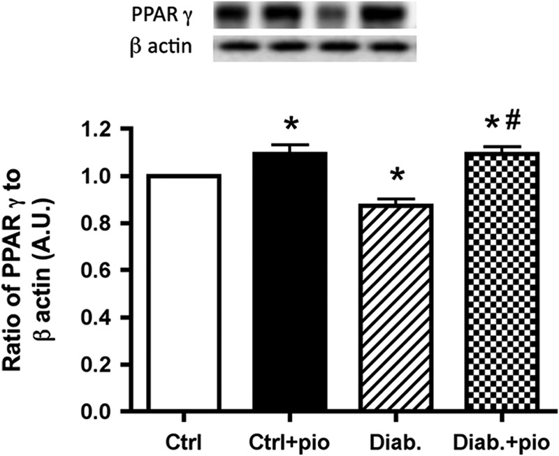
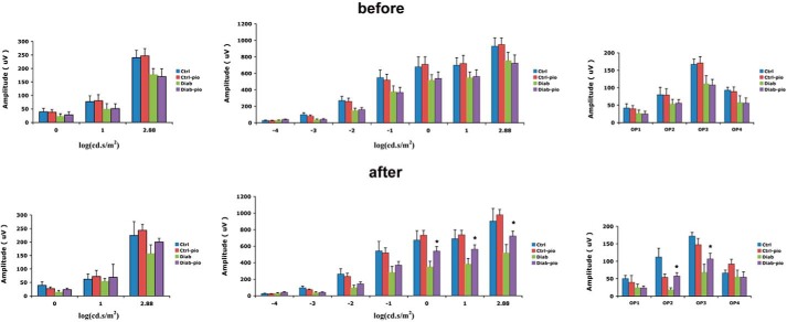
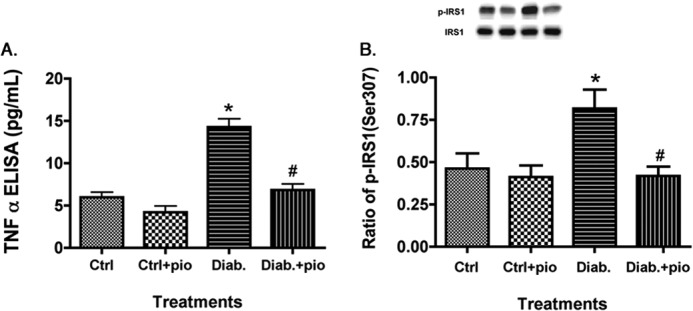
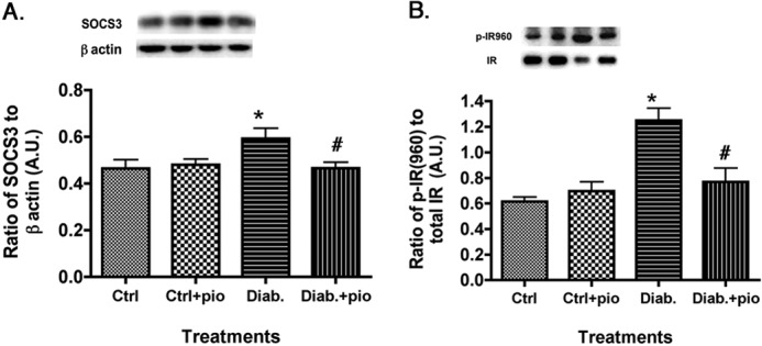
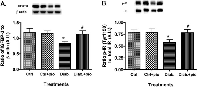
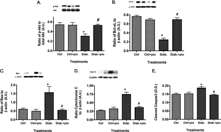
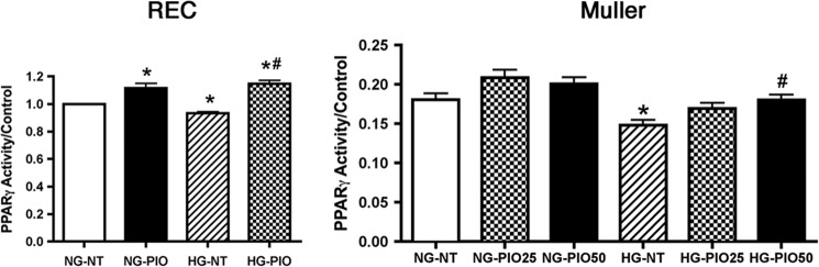
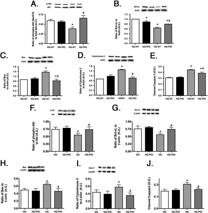
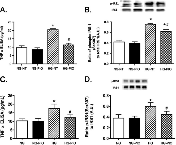
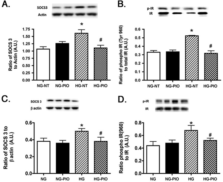
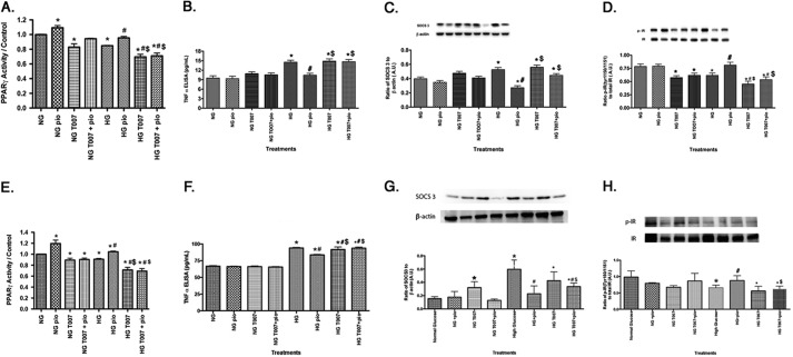
Dysfunctional insulin signaling is a key component of type 2 diabetes. Little is understood of the effects of systemic diabetes on retinal insulin signaling. A number of agents are used to treat patients with type 2 diabetes to normalize glucose levels and improve insulin signaling; however, little has been done to investigate the effects of these agents on retinal insulin signal transduction. We hypothesized that pioglitazone, a peroxisome proliferator-activated receptor γ (PPARγ) agonist, would normalize retinal insulin signal transduction through reduced tumor necrosis factor α (TNFα) and suppressor of cytokine signaling 3 (SOCS3) activities in whole retina and retinal endothelial cells (REC) and Müller cells. To test this hypothesis, we used the BBZDR/Wor type 2 diabetic rat model, as well as REC and Müller cells cultured in normoglycemia and hyperglycemic conditions, to investigate the effects of pioglitazone on TNFα, SOCS3, and downstream insulin signal transduction proteins. We also evaluated pioglitazone's effects on retinal function using electroretinogram and markers of apoptosis. Data demonstrate that 2 months of pioglitazone significantly increased electroretinogram amplitudes in type 2 diabetic obese rats, which was associated with improved insulin receptor activation. These changes occurred in both REC and Müller cells treated with pioglitazone, suggesting that these two cell types are key to insulin resistance in the retina. Taken together, these data provide evidence of impaired insulin signaling in type 2 diabetes rats, which was improved by increasing PPARγ activity. Further investigations of PPARγ actions in the retina may provide improved treatment options.

Keywords: Diabetic Retinopathy; Insulin Receptor; Insulin Receptor Substrate 1 (IRS-1); Insulin Resistance; Suppressor of Cytokine Signaling 3 (SOCS3); Tumor Necrosis Factor (TNF); Type 2 Diabetes.


Figures

FIGURE 1.
FIGURE 1.
Pioglitazone increased PPARγ activity in whole retinal lysates of type 2 diabetic rats. Western blot results from whole retina of lean (Ctrl), lean + pioglitazone (Ctrl+pio), BBZDR/Wor obese (Diab), and BBZDR/Wor obese + pioglitazone (Diab+pio) mice. *, p < 0.05 versus control; #, p < 0.05 versus diabetic. Data are mean ± S.E. (error bars); n = 5 for each group. A.U., arbitrary units.
FIGURE 2.
FIGURE 2.
B-wave amplitudes are improved in diabetic rats treated with pioglitazone. Top, ERG amplitudes for A-wave (left), B-wave (middle), and oscillatory potentials (right) before treatment was started. Bottom, effects of 2 months of pioglitazone treatment. *, p < 0.05 versus diabetic. Data are mean ± S.D. (error bars); n = 5 in all groups.
FIGURE 3.
FIGURE 3.
Pioglitazone reduced TNFα and IRS-1Ser-307 in type 2 diabetic rats. For both A and B, data presented include whole retinal analyses of lean (Ctrl), lean + pioglitazone (Ctrl+pio), BBZDR/Wor obese (Diab), and BBZDR/Wor obese + pioglitazone (Diab+pio) animals. A, ELISA results for TNFα; B, Western blot results for IRS-1Ser-307. *, p < 0.05 versus control; #, p < 0.05 versus diabetic. Data are mean ± S.E. (error bars); n = 5 in all groups.
FIGURE 4.
FIGURE 4.
SOCS3 and IRTyr-960 are reduced in pioglitazone-treated type 2 diabetic rats. For both A and B, data presented include whole retinal analyses of lean (Ctrl), lean + pioglitazone (Ctrl+pio), BBZDR/Wor obese (Diab), and BBZDR/Wor obese + pioglitazone (Diab+pio) animals. A, Western blot results for SOCS3; B, Western blot results for IRTyr-960. *, p < 0.05 versus control; #, p < 0.05 versus diabetic. Data are mean ± S.E. (error bars); n = 5 in all groups. A.U., arbitrary units.
FIGURE 5.
FIGURE 5.
IGFBP-3 and phosphorylation of insulin receptor increased after pioglitazone treatment. For both A and B, data presented include whole retinal analyses of lean (Ctrl), lean + pioglitazone (Ctrl+pio), BBZDR/Wor obese (Diab), and BBZDR/Wor obese + pioglitazone (Diab+pio) animals. A, Western blot results for IGFBP-3; B, Western blot results for IRTyr-1150/1151. *, p < 0.05 versus control; #, p < 0.05 versus diabetic. Data are mean ± S.E.; n = 5 in all groups.A.U., arbitrary units.
FIGURE 6.
FIGURE 6.
Apoptotic markers are reduced in type 2 diabetic rats treated with pioglitazone. For both A and B, data presented include whole retinal analyses of lean (Ctrl), lean + pioglitazone (Ctrl+pio), BBZDR/Wor obese (Diab), and BBZDR/Wor obese + pioglitazone (Diab+pio) animals. A and B show Western blot results for the anti-apoptotic proteins (Akt and Bcl-xL). C–E, Western blot results for pro-apoptotic proteins (Bax, cytochrome c, and cleaved caspase 3). *, p < 0.05 versus control; #, p < 0.05 versus diabetic. Data are mean ± S.E. n = 5 in all groups. A.U., arbitrary units.
FIGURE 7.
FIGURE 7.
PPARγ activity is increased in REC and Müller cells after pioglitazone. For both REC and Müller cells, cells were grown in normal glucose (NG) or high glucose (HG). For REC, cells were treated with 25 μm pioglitazone (PIO), whereas Müller cells were untreated (NT) or treated with 25 or 50 μm pioglitazone. *, p < 0.05 versus normal glucose untreated; #, p < 0.05 versus high glucose untreated. Data are mean ± S.E. n = 4 for all treatments.
FIGURE 8.
FIGURE 8.
Proapoptotic proteins are reduced in REC and Müller cells treated with pioglitazone. All work was done in REC and Müller cells left untreated (NT) or treated with pioglitazone. A–E, data in REC; F–J, Müller cell data. Data show that pioglitazone (PIO) reduced proapoptotic proteins (C–E and H–J) in both cell types. *, p < 0.05 versus normal glucose (NG) untreated; #, p < 0.05 versus high glucose (HG) untreated. Data are mean ± S.E. (error bars); n = 4 for all treatments. A.U., arbitrary units.
FIGURE 9.
FIGURE 9.
TNFα and IRS-1Ser-307 are reduced in both REC and Müller cells after pioglitazone treatment. All work was done in REC and Müller cells left untreated (NT) or treated with pioglitazone (PIO). A and B, data in REC; C and D, Müller cell data. Data show that pioglitazone reduced TNFα and IRS-1Ser-307 in both cell types. *, p < 0.05 versus normal glucose (NG) untreated; #, p < 0.05 versus high glucose (HG) untreated. Data are mean ± S.E. (error bars); n = 4 for all treatments. A.U., arbitrary units.
FIGURE 10.
FIGURE 10.
SOCS3 and IRTyr-960 are reduced by pioglitazone. All work was done in REC and Müller cells left untreated (NT) or treated with pioglitazone. A and B, data in REC; C and D, Müller cell data. Data show that pioglitazone (PIO) reduced SOCS3 and IRTyr-960 in both cell types. *, p < 0.05 versus normal glucose (NG) untreated; #, p < 0.05 versus high glucose (HG) untreated. Data are mean ± S.E. (error bars); n = 4 for all treatments. A.U., arbitrary units.
FIGURE 11.
FIGURE 11.
PPARγ antagonist T0070907 (50 nm) blocks pioglitazone actions on TNFα, SOCS3, and IRTyr-1150/1151. All work was done in REC and Müller cells left untreated (NT), treated with pioglitazone (PIO), treated with T0070907 only, or treated with pioglitazone + T0070907. A–D, REC results; E–H, Müller cell results. A and E show that T0070907 inhibited pioglitazone actions using the PPARγ activity assay. B and F demonstrate that T00709707 antagonized pioglitazone actions on TNFα, whereas C and G show T0070907 actions on SOCS3. D and H show that pioglitazone's ability to increase IRTyr-1150/1151 requires PPARγ because it was blocked by T0070907. *, p < 0.05 versus normal glucose (NG) untreated; #, p < 0.05 versus high glucose untreated; $, p < 0.05 versus high glucose (HG) + pioglitazone. Data are mean ± S.E.; n = 4 for all treatments. A.U., arbitrary units.

References

    1. Ng M., Fleming T., Robinson M., Thomson B., Graetz N., Margono C., Mullany E. C., Biryukov S., Abbafati C., Abera S. F., Abraham J. P., Abu-Rmeileh N. M., Achoki T., AlBuhairan F. S., Alemu Z. A., Alfonso R., Ali M. K., Ali R., Guzman N. A., Ammar W., Anwari P., Banerjee A., Barquera S., Basu S., Bennett D. A., Bhutta Z., Blore J., Cabral N., Nonato I. C., Chang J. C., Chowdhury R., Courville K. J., Criqui M. H., Cundiff D. K., Dabhadkar K. C., Dandona L., Davis A., Dayama A., Dharmaratne S. D., Ding E. L., Durrani A. M., Esteghamati A., Farzadfar F., Fay D. F., Feigin V. L., Flaxman A., Forouzanfar M. H., Goto A., Green M. A., Gupta R., Hafezi-Nejad N., Hankey G. J., Harewood H. C., Havmoeller R., Hay S., Hernandez L., Husseini A., Idrisov B. T., Ikeda N., Islami F., Jahangir E., Jassal S. K., Jee S. H., Jeffreys M., Jonas J. B., Kabagambe E. K., Khalifa S. E., Kengne A. P., Khader Y. S., Khang Y. H., Kim D., Kimokoti R. W., Kinge J. M., Kokubo Y., Kosen S., Kwan G., Lai T., Leinsalu M., Li Y., Liang X., Liu S., Logroscino G., Lotufo P. A., Lu Y., Ma J., Mainoo N. K., Mensah G. A., Merriman T. R., Mokdad A. H., Moschandreas J., Naghavi M., Naheed A., Nand D., Narayan K. M., Nelson E. L., Neuhouser M. L., Nisar M. I., Ohkubo T., Oti S. O., Pedroza A., Prabhakaran D., Roy N., Sampson U., Seo H., Sepanlou S. G., Shibuya K., Shiri R., Shiue I., Singh G. M., Singh J. A., Skirbekk V., Stapelberg N. J., Sturua L., Sykes B. L., Tobias M., Tran B. X., Trasande L., Toyoshima H., van de Vijver S., Vasankari T. J., Veerman J. L., Velasquez-Melendez G., Vlassov V. V., Vollset S. E., Vos T., Wang C., Wang S. X., Weiderpass E., Werdecker A., Wright J. L., Yang Y. C., Yatsuya H., Yoon J., Yoon S. J., Zhao Y., Zhou M., Zhu S., Lopez A. D., Murray C. J., Gakidou E. (2014) Global, regional, and national prevalence of overweight and obesity in children and adults during 1980–2013: a systematic analysis for the Global Burden of Disease Study 2013. Lancet 10.1016/S0140-6736(14)60460-8 - DOI - PMC - PubMed
    1. Tawfik A., Sanders T., Kahook K., Akeel S., Elmarakby A., Al-Shabrawey M. (2009) Suppression of retinal peroxisome proliferator-activated receptor γ in experimental diabetes and oxygen-induced retinopathy: role of NADPH oxidase. Invest. Ophthalmol. Vis. Sci. 50, 878–884 - PubMed
    1. Malchiodi-Albedi F., Matteucci A., Bernardo A., Minghetti L. (2008) PPAR-γ, Microglial cells, and ocular inflammation: new venues for potential therapeutic approaches. PPAR Res. 2008, 295784. - PMC - PubMed
    1. Yau H., Rivera K., Lomonaco R., Cusi K. (2013) The future of thiazolidinedione therapy in the management of type 2 diabetes mellitus. Curr. Diab. Rep. 13, 329–341 - PubMed
    1. Iwata M., Haruta T., Usui I., Takata Y., Takano A., Uno T., Kawahara J., Ueno E., Sasaoka T., Ishibashi O., Kobayashi M. (2001) Pioglitazone ameliorates tumor necrosis factor-α-induced insulin resistance by a mechanism independent of adipogenic activity of peroxisome proliferator-activated receptor-γ. Diabetes 50, 1083–1092 - PubMed

Publication types

MeSH terms