Review
. 2014 Nov:96:38-45.
doi: 10.1016/j.mvr.2014.07.008. Epub 2014 Jul 30.

Primary and secondary lymphatic valve development: molecular, functional and mechanical insights

Affiliations
Review

Primary and secondary lymphatic valve development: molecular, functional and mechanical insights

Eleni Bazigou et al. Microvasc Res. 2014 Nov.

Abstract

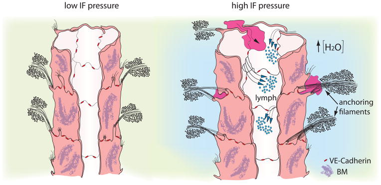
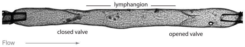
Fluid homeostasis in vertebrates critically relies on the lymphatic system forming a hierarchical network of lymphatic capillaries and collecting lymphatics, for the efficient drainage and transport of extravasated fluid back to the cardiovascular system. Blind-ended lymphatic capillaries employ specialized junctions and anchoring filaments to encourage a unidirectional flow of the interstitial fluid into the initial lymphatic vessels, whereas collecting lymphatics are responsible for the active propulsion of the lymph to the venous circulation via the combined action of lymphatic muscle cells and intraluminal valves. Here we describe recent findings on molecular and physical factors regulating the development and maturation of these two types of valves and examine their role in tissue-fluid homeostasis.

Keywords: Biomechanics; Endothelium; Interstitial fluid; Lymphatic vessel; Valve.


Conflict of interest statement

CONFLICT OF INTEREST

There are no conflicts of interest to state.

Figures

Figure 1
Figure 1
Primary valves in initial lymphatics respond to fluid pressure changes and allow drainage of IF and passage of cells. When IF pressure is low (left), lymph flow is not promoted towards the lumen. Upon increase in IF pressure (right) anchoring filaments pull the overlapping junctions apart and allow entry of fluid (lymph) and dendritic cells (purple) towards the lumen. The integrity of the lumen is secured by VE-Cadherin located at the “buttons” (red) [Adapted from Leak and Burke, 1968, (Leak and Burke, 1968)].
Figure 2
Figure 2
Lymphangion is the functional unit of collecting lymphatic vessels. Image of a cannulated collecting lymphatic vessel containing two intraluminal valves, the first one is closed and the lymph is transported through the second open valve to the next lymphangion. Objects at the far left and right portions of the vessel are cannulating micropipettes. Calibration bar = 100 μm (Image kindly provided by Dr. Michael J. Davis, University of Missouri).
Figure 3
Figure 3
Flow and NO transport simulations performed using a computational geometry constructed from confocal images of a rat mesenteric lymphatic vessel. The direction of flow is from bottom to top. Left) Distributions of normalized NO production (prescribed to increase with shear stress) at the wall. The areas of lowest NO production occur in the regions adjacent to the valve leaflets encapsulated by the sinus. Right) Distributions of normalized NO concentration at the wall of the vessel overlaid with representative lymph flow velocity streamlines. NO concentration values are normalized by a characteristic inlet concentration, Co. While streamlines adjacent to the valve leaflets appear vortex-like in nature, these are essentially regions of flow stagnation [Images based on data from Wilson et al. (Wilson et al., 2013)].
Figure 4
Figure 4
Development of intraluminal lymphatic valves. (A) Schematic of a phenocopied embryonic lymphatic vessel undergoing remodeling in steps 1. Establishment of valve territory, 2. LEC reorientation and active migration at ridges of lymphatic vessels, 3. ECM organization in a fibrous ring and 4. Valve leaflet elongation. (B) Assumption of the same vessel in its mature form as a collecting lymphatic vessel showing fully developed intraluminal valves and LMCs.

References

    1. Abtahian F, et al. Regulation of blood and lymphatic vascular separation by signaling proteins SLP-76 and Syk. Science. 2003;299:247–251. - PMC - PubMed
    1. Aukland K, Nicolaysen G. Interstitial fluid volume: local regulatory mechanisms. Physiological reviews. 1981;61:556–643. - PubMed
    1. Aukland K, Reed R. Interstitial-lymphatic mechanisms in the control of extracellular fluid volume. Physiological reviews. 1993;73:1–78. - PubMed
    1. Baluk P, et al. Functionally specialized junctions between endothelial cells of lymphatic vessels. The Journal of experimental medicine. 2007;204:2349–2362. - PMC - PubMed
    1. Bank RA, et al. The increased swelling and instantaneous deformation of osteoarthritic cartilage is highly correlated with collagen degradation. Arthritis & Rheumatism. 2000;43:2202–2210. - PubMed

Publication types