

Parental somatic mosaicism is underrecognized and influences recurrence risk of genomic disorders
- PMID: 25087610
- PMCID: PMC4129404
- DOI: 10.1016/j.ajhg.2014.07.003
Parental somatic mosaicism is underrecognized and influences recurrence risk of genomic disorders
Abstract
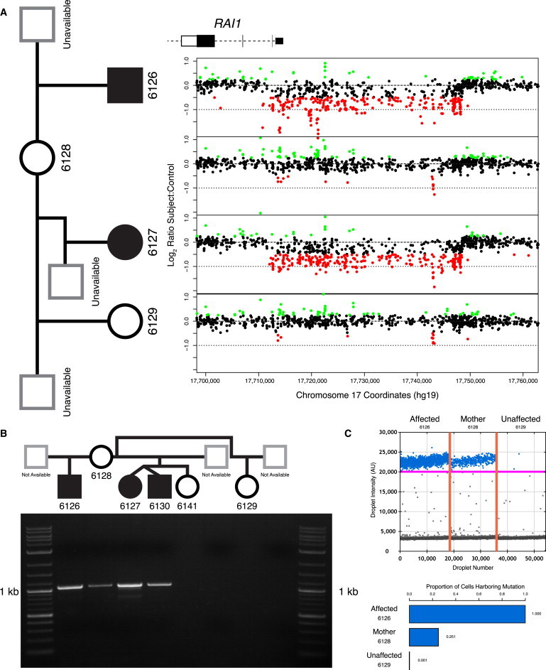
New human mutations are thought to originate in germ cells, thus making a recurrence of the same mutation in a sibling exceedingly rare. However, increasing sensitivity of genomic technologies has anecdotally revealed mosaicism for mutations in somatic tissues of apparently healthy parents. Such somatically mosaic parents might also have germline mosaicism that can potentially cause unexpected intergenerational recurrences. Here, we show that somatic mosaicism for transmitted mutations among parents of children with simplex genetic disease is more common than currently appreciated. Using the sensitivity of individual-specific breakpoint PCR, we prospectively screened 100 families with children affected by genomic disorders due to rare deletion copy-number variants (CNVs) determined to be de novo by clinical analysis of parental DNA. Surprisingly, we identified four cases of low-level somatic mosaicism for the transmitted CNV in DNA isolated from parental blood. Integrated probabilistic modeling of gametogenesis developed in response to our observations predicts that mutations in parental blood increase recurrence risk substantially more than parental mutations confined to the germline. Moreover, despite the fact that maternally transmitted mutations are the minority of alleles, our model suggests that sexual dimorphisms in gametogenesis result in a greater proportion of somatically mosaic transmitting mothers who are thus at increased risk of recurrence. Therefore, somatic mosaicism together with sexual differences in gametogenesis might explain a considerable fraction of unexpected recurrences of X-linked recessive disease. Overall, our results underscore an important role for somatic mosaicism and mitotic replicative mutational mechanisms in transmission genetics.
Copyright © 2014 The American Society of Human Genetics. Published by Elsevier Inc. All rights reserved.
Figures


Comment in
-
Somatic mosaicism in parents may cause single-gene disorders in children: latest study calculates incidence of parental mosaicism higher than previous research suggests.Am J Med Genet A. 2014 Nov;164A(11):viii-ix. doi: 10.1002/ajmg.a.36818. Am J Med Genet A. 2014. PMID: 25327470 No abstract available.
References
-
- Bakker E., Van Broeckhoven C., Bonten E.J., van de Vooren M.J., Veenema H., Van Hul W., Van Ommen G.J., Vandenberghe A., Pearson P.L. Germline mosaicism and Duchenne muscular dystrophy mutations. Nature. 1987;329:554–556. - PubMed
-
- Seshadri R., Kutlaca R.J., Trainor K., Matthews C., Morley A.A. Mutation rate of normal and malignant human lymphocytes. Cancer Res. 1987;47:407–409. - PubMed
-
- Erickson R.P. Somatic gene mutation and human disease other than cancer: an update. Mutat. Res. 2010;705:96–106. - PubMed
-
- Lupski J.R. Genetics. Genome mosaicism—one human, multiple genomes. Science. 2013;341:358–359. - PubMed
Publication types
MeSH terms
Grants and funding
LinkOut - more resources
Full Text Sources
Other Literature Sources
Medical
