. 2014 Aug 7;95(2):218-26.
doi: 10.1016/j.ajhg.2014.07.004. Epub 2014 Jul 31.

SPEG interacts with myotubularin, and its deficiency causes centronuclear myopathy with dilated cardiomyopathy

Affiliations

SPEG interacts with myotubularin, and its deficiency causes centronuclear myopathy with dilated cardiomyopathy

Pankaj B Agrawal et al. Am J Hum Genet. .

Abstract

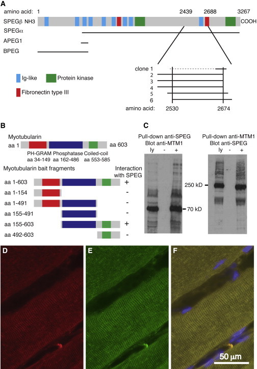
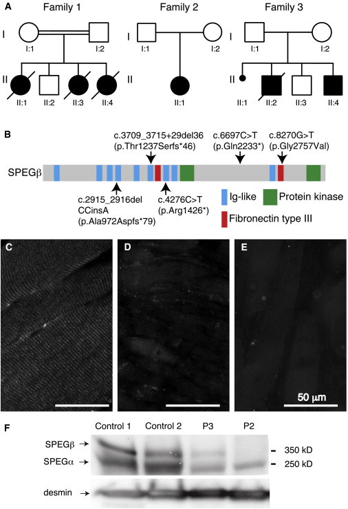
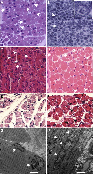
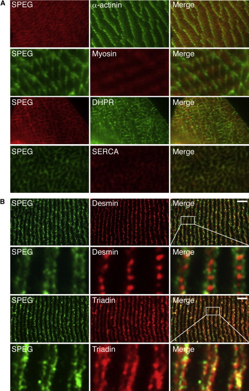
Centronuclear myopathies (CNMs) are characterized by muscle weakness and increased numbers of central nuclei within myofibers. X-linked myotubular myopathy, the most common severe form of CNM, is caused by mutations in MTM1, encoding myotubularin (MTM1), a lipid phosphatase. To increase our understanding of MTM1 function, we conducted a yeast two-hybrid screen to identify MTM1-interacting proteins. Striated muscle preferentially expressed protein kinase (SPEG), the product of SPEG complex locus (SPEG), was identified as an MTM1-interacting protein, confirmed by immunoprecipitation and immunofluorescence studies. SPEG knockout has been previously associated with severe dilated cardiomyopathy in a mouse model. Using whole-exome sequencing, we identified three unrelated CNM-affected probands, including two with documented dilated cardiomyopathy, carrying homozygous or compound-heterozygous SPEG mutations. SPEG was markedly reduced or absent in two individuals whose muscle was available for immunofluorescence and immunoblot studies. Examination of muscle samples from Speg-knockout mice revealed an increased frequency of central nuclei, as seen in human subjects. SPEG localizes in a double line, flanking desmin over the Z lines, and is apparently in alignment with the terminal cisternae of the sarcoplasmic reticulum. Examination of human and murine MTM1-deficient muscles revealed similar abnormalities in staining patterns for both desmin and SPEG. Our results suggest that mutations in SPEG, encoding SPEG, cause a CNM phenotype as a result of its interaction with MTM1. SPEG is present in cardiac muscle, where it plays a critical role; therefore, individuals with SPEG mutations additionally present with dilated cardiomyopathy.


Figures

Figure 1
Figure 1
SPEG Interacts with MTM1 (A) Schematic of the six different SPEG Y2H prey clones that interacted with full-length MTM1 bait; they are lined up under the four alternative SPEG transcripts (indicated on the left). The broken line indicates that the 5′ end of clone 1 was unknown. (B) A schematic of MTM1 illustrates the location of three major domains—PH-GRAM (amino acids 34–149), phosphatase (amino acids 162–486), and coiled coil (amino acids 553–585)—above the map of fragments used for deletion mapping of the region responsible for interactions with SPEG. Deletion analysis of MTM1 showed that the phosphatase and coiled-coil domains together were necessary to mediate the interaction with SPEG. (C) SPEG and MTM1 coimmunoprecipitated from C2C12 myotube lysates with the use of rabbit anti-SPEG generated against a FLAG-tagged APEG-1 fusion protein and anti-MTM1 antibodies (r1947). Abbreviations are as follows: ly, total cell lysates; −, no precipitating antibody; and +, immunoprecipitated with “pull-down” antisera (indicated at the top) prior to gel electrophoresis and immunoblotting with indicated antisera. (D–F) Indirect immunofluorescence analysis of SPEG (red, D), MTM1 (green, E), and a merged image, including blue DAPI-stained nuclei (F), with the use of rabbit anti-SPEG and mouse anti-MTM1 (1:80, HPA010008, Sigma-Aldrich) antibodies revealed their colocalization in human skeletal muscle. The scale bar represents 50 μm.
Figure 2
Figure 2
Genetic and Molecular Findings in Three Families Affected by SPEG Mutations (A) Pedigree of the three families carrying SPEG mutations. Family 1 was consanguineous, whereas families 2 and 3 were not. The probands were II:4 (P1) for family 1, II:1 (P2) for family 2, and II:4 (P3) for family 3. (B) Distribution of alterations across the schematic of SPEG. Domains are also indicated. (C–E) Indirect immunofluorescence analysis using rabbit anti-SPEG antibody (NBP1-90134, Novus Biologicals) in muscle-biopsy specimens from a representative age-matched human control individual (C) and probands P2 (II:4 from family 2, D) and P3 (II:4 from family 3, E) showed a loss of the striated pattern and a marked and reproducible reduction of overall SPEG staining in muscle from both subjects. (F) Immunoblot analysis using rabbit anti-SPEG in muscle-biopsy specimens from two unaffected control individuals and subjects 2 and 3. Restaining the filter with anti-desmin antibodies (1466-1, Epitomics) confirmed adequate loading of lanes and demonstrated robust levels of desmin in these muscles.
Figure 3
Figure 3
Histopathological Findings in Human Subjects and a Mouse Model of SPEG Deficiency (A–D) Light microscopic findings in muscle-biopsy specimens from human probands P1 (A and B), P2 (II:4 from family 2 in Figure 2, C), and P3 (II:4 from family 3 in Figure 2, D) included increased central nuclei on hematoxylin and eosin (H&E) staining (arrows in A, C, and D) and subsarcolemmal ringed and central dense areas, also called necklace fibers (arrows and inset, with NADH tetrazolium reductase staining, in B). (E and F) Histopathological and ultrastructural findings in skeletal muscles from SPEG-deficient and wild-type (WT) littermate mice. H&E staining of paraspinal muscles revealed higher numbers of central nuclei (arrows) in 1-day-old SPEG-deficient mice (F) than in WT controls (E). (G and H) Transmission electron microscopic findings in skeletal-muscle (quadriceps) specimens obtained from Speg-KO (H) and WT littermate (G) mice. Several centrally placed nuclei were present in the specimen from a Speg-KO mouse (arrows, H), and in comparison, peripherally located nuclei were seen in the control mouse (arrows, G). Scale bars represent 4 μm.
Figure 4
Figure 4
SPEG Localization in Normal Skeletal Muscle (A) Ultrathin sections from frozen WT mouse quadriceps muscle were coimmunostained with mouse monoclonal anti-α-actinin-2 (clone EA-53, Sigma), mouse monoclonal anti-myosin heavy chain fast (clone MY-32, Sigma), mouse monoclonal anti-dihydropyridine receptor (DHPR, clone D218, Sigma), mouse monoclonal anti-sarcoplasmic reticulum Ca2+ATPase (SERCA, clone IIH11, Sigma), and rabbit anti-SPEG (indicated). As seen in the merged images, SPEG staining partially overlapped with α-actinin depending on the plane of section and did not colocalize with myosin but did colocalize with both DHPR and SERCA. (B) Longitudinal sections from frozen human quadriceps muscles were coimmunostained with monoclonal mouse anti-human desmin (clone D33, Dako), mouse monoclonal anti-rabbit triadin (clone GE 4.90, Abcam), and rabbit anti-SPEG (indicated). SPEG appeared as a doublet over the myofibrils and flanking desmin, which is found between Z lines. SPEG largely colocalized with triadin, which is found at the junctional SR. Scale bars represent 7 μm.
Figure 5
Figure 5
Analysis of SPEG and Desmin Distribution in MTM1-Deficient Skeletal Muscles Representative indirect immunofluorescence images for SPEG (rabbit anti-SPEG, green) and desmin (clone D33, red) in skeletal-muscle specimens from an unaffected control individual (top row), a boy with X-linked myotubular myopathy due to an MTM1 mutation (second row), a WT mouse (third row), and an Mtm1-KO mouse (bottom row). Merged images, including nuclei stained blue, are shown on the right. Note the abnormal clumping and accumulations of colocalized SPEG and desmin in the MTM1-deficient muscles. The scale bar represents 100 μm.

References

    1. Nance J.R., Dowling J.J., Gibbs E.M., Bönnemann C.G. Congenital myopathies: an update. Curr. Neurol. Neurosci. Rep. 2012;12:165–174. - PMC - PubMed
    1. Pierson C.R., Tomczak K., Agrawal P., Moghadaszadeh B., Beggs A.H. X-linked myotubular and centronuclear myopathies. J. Neuropathol. Exp. Neurol. 2005;64:555–564. - PubMed
    1. Romero N.B., Bitoun M. Centronuclear myopathies. Semin. Pediatr. Neurol. 2011;18:250–256. - PubMed
    1. Laporte J., Hu L.J., Kretz C., Mandel J.L., Kioschis P., Coy J.F., Klauck S.M., Poustka A., Dahl N. A gene mutated in X-linked myotubular myopathy defines a new putative tyrosine phosphatase family conserved in yeast. Nat. Genet. 1996;13:175–182. - PubMed
    1. Bitoun M., Maugenre S., Jeannet P.Y., Lacène E., Ferrer X., Laforêt P., Martin J.J., Laporte J., Lochmüller H., Beggs A.H. Mutations in dynamin 2 cause dominant centronuclear myopathy. Nat. Genet. 2005;37:1207–1209. - PubMed

Publication types

MeSH terms

Substances