. 2014 Nov;32(11):1416-23.
doi: 10.1002/jor.22697. Epub 2014 Aug 2.

Early detection of heterotopic ossification using near-infrared optical imaging reveals dynamic turnover and progression of mineralization following Achilles tenotomy and burn injury

Affiliations

Early detection of heterotopic ossification using near-infrared optical imaging reveals dynamic turnover and progression of mineralization following Achilles tenotomy and burn injury

Joseph E Perosky et al. J Orthop Res. 2014 Nov.

Abstract

Heterotopic ossification (HO) is the abnormal formation of bone in soft tissue. Current diagnostics have low sensitivity or specificity to incremental progression of mineralization, especially at early time points. Without accurate and reliable early diagnosis and intervention, HO progression often results in incapacitating conditions of limited range of motion, nerve entrapment, and pain. We hypothesized that non-invasive near-infrared (NIR) optical imaging can detect HO at early time points and monitor heterotopic bone turnover longitudinally. C57BL6 mice received an Achilles tenotomy on their left hind limb in combination with a dorsal burn or sham procedure. A calcium-chelating tetracycline derivative (IRDye 680RD BoneTag) was injected bi-weekly and imaged via NIR to measure accumulative fluorescence for 11 wk and compared to in vivo microCT images. Percent retention of fluorescence was calculated longitudinally to assess temporal bone resorption. NIR detected HO as early as five days and revealed a temporal response in HO formation and turnover. MicroCT could not detect HO until 5 wk. Confocal microscopy confirmed fluorophore localization to areas of HO. These findings demonstrate the ability of a near-infrared optical imaging strategy to accurately and reliably detect and monitor HO in a murine model.

Keywords: biomarkers; heterotopic Ossification; microCT; molecular imaging.


Figures

Figure 1
Figure 1
Time course of BoneTag distribution at the proximal tibia. Mice injected with BoneTag680 or a carboxylated control probe demonstrated early vascular perfusion of both imaging agents. BoneTag680 was retained specifically at skeletal sites, while the control probe was rapidly cleared through the vasculature after 5 min and virtually absent at 24 h. Inset: Probe fluorescence over initial 2 h magnified for clarity.
Figure 2
Figure 2
(A) Accumulative fluorescence in limbs subject to Achilles tenotomy was greater than contralateral controls in both (A) 30° and (B) 60° burn groups (* p < 0.05 tenotomy vs. control). In both tenotomy groups, accumulative fluorescence significantly increased in a temporal manner. (B) Burn significantly accelerated HO by 5 wk over tenotomy alone (# p < 0.05 tenotomy + burn (B) versus tenotomy (A) with a trend toward continuing differences at 7, 9, and 11 wk (p < 0.10). (C) Heterotopic ossification was not detected by microCT until 5 wk. (D) Once detected, microCT was highly predictive of accumulative fluorescence; R2 = 0.63, p < 0.0005.
Figure 3
Figure 3
Temporal progression of heterotopic ossification was visualized by NIR imaging for (A) 30° tenotomy limbs and (B) tenotomy plus burn limbs. (C) Intact contralateral limbs from the burn group, which did not form HO are shown for comparison.
Figure 4
Figure 4
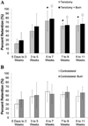
(A) Percent retention of fluorescence was significantly lower from 5 days to 3 wk compared to the 5–7, 7–9, and 9–11 wk time points (*, ◇ = p < 0.05 for tenotomized and tenotomised + burn groups respectively), indicating increased localized resorptive activity in both the tenotomy and tenotomy + burn groups until 3 wk, which normalizes by 5 wk. (B) This is in contrast to the contralateral limbs of both groups, which did not show any changes in fluorophore retention over time course.
Figure 5
Figure 5
Ex vivo near-infrared (A) and microCT (B) images taken at 11 wk confirm the formation of mid-tibial HO, as well as HO formed around and on top of the posterior calcaneus. Formation of HO in the mid-tibia as well as the posterior-calcaneus was observed in all mice by cessation of observation at 11 wk time point.
Figure 6
Figure 6
(A) confocal microscopy of mid-tibia region confirmed probe binding in regions of HO (B) as well as pre-existing cortical bone (D). Subsequent staining with toluidine blue confirmed HE (C) and cortical bone (E) phenotype. Confocal image of posterior calcaneous and corresponding (G) toluidine blue-stained section. (H,I): Unorganized HO formed on top of well-organized cortical bone (J,K). Scale bar = 100 µm.

References

    1. Nauth A, Giles E, Potter BK, et al. Heterotopic ossification in orthopaedic trauma. J Orthop Trauma. 2012;26:684–688. - PMC - PubMed
    1. Vanden Bossche L, Vanderstraeten G. Heterotopic ossification: a review. J Rehabil Med. 2005;37:129–136. - PubMed
    1. Blokhuis TJ, Frölke JP. Is radiation superior to indomethacin to prevent heterotopic ossification in acetabular fractures? a systematic review. Clin Orthop Relat Res. 2009;467:526–530. - PMC - PubMed
    1. Burd TA, Lowry KJ, Anglen JO. Indomethacin compared with localized irradiation for the prevention of heterotopic ossification following surgical treatment of acetabular fractures. J Bone Joint Surg Am. 2001;83:1783–1788. - PubMed
    1. Cullen N, Perera J. Heterotopic Ossification: pharmacologic options. J Head Trauma Rehabil. 2009;24:69–71. - PubMed

Publication types

MeSH terms