. 2014 Sep 4;55(5):758-70.
doi: 10.1016/j.molcel.2014.06.032. Epub 2014 Jul 31.

A synthetic biology approach identifies the mammalian UPR RNA ligase RtcB

Affiliations

A synthetic biology approach identifies the mammalian UPR RNA ligase RtcB

Yanyan Lu et al. Mol Cell. .

Abstract

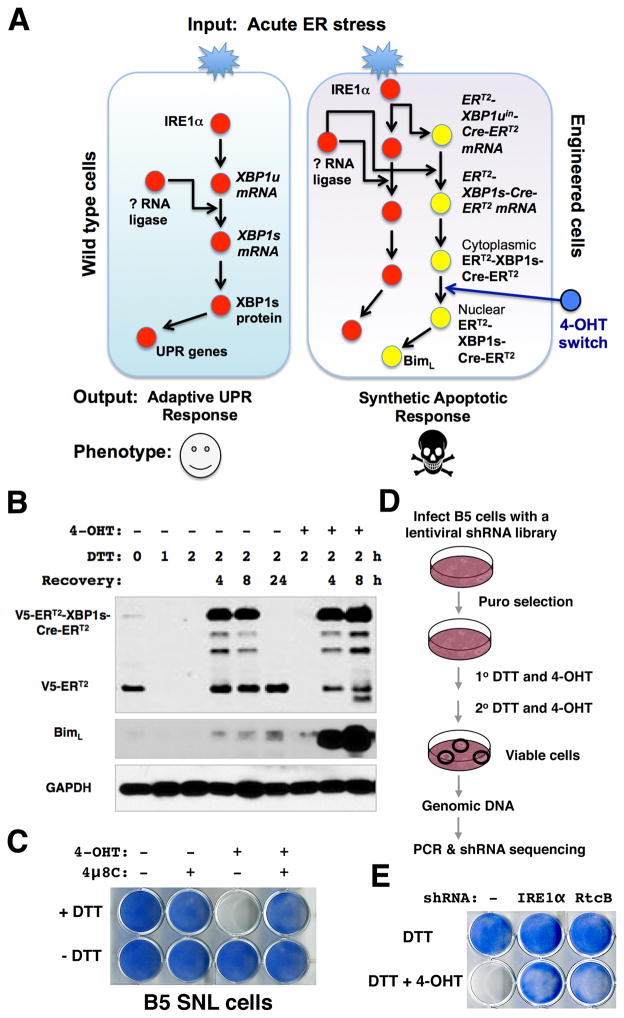
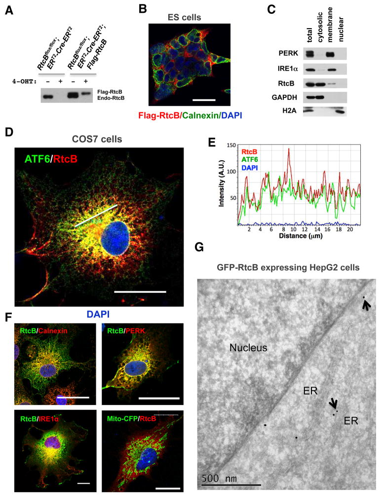
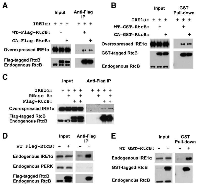
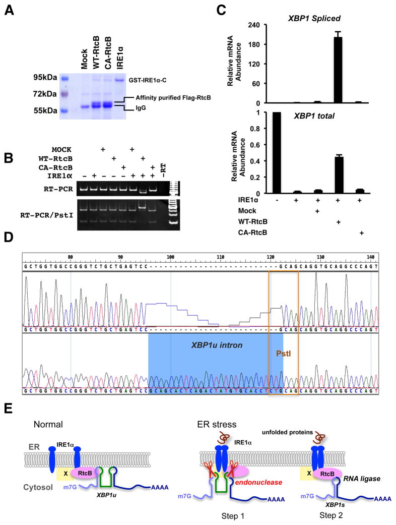
Signaling in the ancestral branch of the unfolded protein response (UPR) is initiated by unconventional splicing of HAC1/XBP1 mRNA during endoplasmic reticulum (ER) stress. In mammals, IRE1α has been known to cleave the XBP1 intron. However, the enzyme responsible for ligation of two XBP1 exons remains unknown. Using an XBP1 splicing-based synthetic circuit, we identify RtcB as the primary UPR RNA ligase. In RtcB knockout cells, XBP1 mRNA splicing is defective during ER stress. Genetic rescue and in vitro splicing show that the RNA ligase activity of RtcB is directly required for the splicing of XBP1 mRNA. Taken together, these data demonstrate that RtcB is the long-sought RNA ligase that catalyzes unconventional RNA splicing during the mammalian UPR.


Figures

Figure 1
Figure 1. A synthetic circuit identifies RtcB as a candidate UPR RNA ligase
A. A synthetic genetic circuit creates an apoptotic phenotype in response to acute ER stress. By coupling of an XBP1-splicing dependent Cre sensor with endogenous IRE1α, the 4-OHT-regulated Cre activates Bim expression and triggers a robust apoptotic response in the genetically engineered SNL cells. B. The UPR-induced splicing of V5-ERT2-XBP1uin-Cre- ERT2sensor and the induction of Bim in B5 SNL cells. DTT in combination with 4-OHTinduced an accumulation of full-length V5-ERT2-XBP1s-Cre- ERT2proteins and a robust induction of Bim. Note that the truncated V5-ERT2 proteins were depleted in cells acutely treated with DTT without recovery. The B5 cells underwent massive cell death 24 hours after being incubated with DTT and 4-OHT and the sample was not included in this analysis. C. Images of methylene blue staining showed that a combination of DTT and 4-OHT treatments was sufficient to cause robust apoptosis. Inhibition of the endonuclease activity of IRE1α by 4μ8C completely blocked the synthetic apoptosis. D. Experimental design for a genome-wide RNAi screen to identify genetic suppressors of the synthetic apoptosis in B5 SNL cells. E. RtcB was identified as a candidate UPR RNA ligase. The methylene blue stains showed that unlike parental B5 cells, the B5 cells that stably expressed an shRNA against RtcB were viable after DTT and 4-OHT treatment. IRE1α shRNA served as a positive control.
Figure 2
Figure 2. RtcBcKO cells exhibit a defect in endogenous Tyr pre-tRNA splicing
A. Sequence alignment of all 10 tyrosine pre-tRNA genes from the mouse genome. Primers used in RT-PCR analysis are highlighted. B. Verification of indicated primers for detecting unspliced and spliced Tyr tRNA. Primer S and Primer AS1 predominantly detect unspliced Tyr pre-tRNA in an RT-PCR assay as similar products were amplified from both genomic DNA and cDNA samples. Primer S and Primer As2 specifically detect a 73-bp spliced product only from the cDNA. C. RT-PCR analysis of Tyr tRNA splicing in RtcBcKO ES cells. At day 5 after 4-OHT treatment, the level of spliced Tyr tRNA was decreased in RtcB-depleted cells. D. RT-qPCR analysis of the relative abundance of unspliced Tyr pre-tRNA and spliced Tyr tRNA in RtcBcKO cells. GAPDH was used as an internal control. In comparison with the control, only 30% of spliced Tyr tRNA remained 5 days after 4-OHT induction. E. RT-qPCR analysis of the relative abundance of 4 intronless tRNAs in RtcBcKO cells. F. Quantitative western blot analysis of puromycin-labeled total cellular proteins in RtcBcKO cells. A Coomassieblue stained duplicate gel served as the loading control. Quantification of the intensity of fluorescence signal for each lane was plotted. * p< 0.05, ** p< 0.01, *** p< 0.0001, compared to the control groups.
Figure 3
Figure 3. Unconventional XBP1 mRNA splicing is defective in RtcB deficient cells
A. Western blot analysis showed that the accumulation of XBP1s proteins was blocked almost completely in response to Tg in RtcB deficient cells. Note that under the same condition, IRE1α phosphorylation, PERK phosphorylation and CHOP induction remained intact in RtcB deficient cells. B-C. The splicing of XBP1umRNA is defective in RtcBcKO cells. RT-PCR showed that both spliced and unsplicedXBP1 mRNAs were significantly reduced when RtcB were depleted (B). The accumulation of spliced XBP1s mRNA was blocked by IRE1α specific inhibitor 4μ8C in both control and RtcB-deficient cells (C). D. RT-qPCR analysis showed that the relative abundance of XBP1s mRNA in 4-OHT treated cells was drastically reduced in response to ER stress compared to that in untreated control cells. To induce ER stress, the cells were treated with Tm for 4 h, Tg for 2 h and DTT for 2h, respectively. In RtcB deficient cells, the levels of total XBP1 mRNA was also decreased but to a lesser extent. GAPDH served as an internal control in the experiments. * p< 0.05, ** p< 0.01, *** p< 0.0001, compared to the control groups. E. The splicing of Flag-tagged full-length XBP1u reporter in RtcB deficient cells. A constitutively expressed XBP1ucDNA expression vector was stably transfected into RtcBcKO ES cells, providing a reporter cell line lacking the endogenous XBP1 mediated feedback regulation. In E8H2 cells expressing Flag-XBP1ucDNA, XBP1s protein was not induced by DTT after 4-OHT treatment. Note that RtcB knockout had no direct effect on the expression of XBP1s encoded by a spliced Flag-XBP1scDNA transgene.
Figure 4
Figure 4. Genetic rescue shows that RtcB acts as an RNA ligase for XBP1 splicing
A. The RtcBcKO cells were stably transfected with Flag-RtcBWT or Flag-RtcBC122A. Flag-RtcB proteins were expressed at a level similar to endogenous RtcB. B. Methylene blue stains showed that like RtcB null cells (Fig. S3D), RtcBC122A-expressing ES cells exhibited a severe growth defect after deleting endogenous RtcB. C. A confocal immunofluorescence image showing that GFP-RtcBWT and Flag-RtcBC122A have overlapping subcellular localization in transfected COS7 cells. D. In 4-OHT treated Flag-RtcBC122A-expressing E8H2 cells, ligase-dead RtcB failed to rescue the XBP1u splicing defect. No XBP1s protein was detected after Tm treatment. E–F. Similar to the experiment described in Fig. 3D, RT-qPCR showed that wild type RtcB rescued the splicing defect of XBP1u whereas C122A mutant did not. The basal level of XBP1s was significantly reduced and the induction of XBP1s by Tg was abolished in RtcBC122A -expressing cells. Similarly, under both basal and induced conditions, the levels of total XBP1 mRNA were decreased significantly in RtcBC122A-rescued cells.* p< 0.05, ** p< 0.01, *** p< 0.0001, compared to the control groups.
Figure 5
Figure 5. Subcellular localization of RtcB
A. Expression of Flag-RtcB proteins in genetically rescued RtcBcKO ES cells. B. A confocal imunofluorescence image showing that RtcB was enriched in the perinuclear region in ES cells. C. Cell fractionation showed that a portion of endogenous RtcB was associated with the membrane fraction. IRE1α and PERK are ER transmembrane proteins. GAPDH and Histone H2A are cytosolic and nuclear, respectively. D–F. Subcellular localization of RtcB with the ER resident proteins. COS7 cells were co-transfected with Flag-RtcB and indicated ER markers including GFP-ATF6 (in D), calnexin, IRE1αKA, PERK-ΔC-9E10 and a mitochondria marker mito-CFP (in F). Confocal images showed that RtcB overlapped partially with these ER markers. Note that kinase-dead IRE1α and C-terminal truncated PERK were used in the experiment to achieve higher levels of expression. Bars, 25 μm in B, D and F. In E, an intensity plot along the line drawn in the confocal image showed that RtcB was colocalized with ATF6 in this region. G. An immuno-gold labeled EM image showing that GFP-RtcB proteins were labeled on the ER in HepG2 cells. Gold particles on the ER membrane are marked by arrows.
Figure 6
Figure 6. RtcB is in complex with IRE1α
A. In HEK293T cells stably transfected with either Flag-RtcBWT or Flag-RtcBC122A, the level of Flag-RtcB was comparable to that of endogenous RtcB (lower panel, input). Anti-Flag co-IP showed that both wild type and ligase-dead RtcB proteins were associated with overexpressed IRE1α. Parental HEK293T cells that overexpressed IRE1α were used as a negative control. B. Using glutathione-Sepharose pull-down, a similar experiment was performed with GST-RtcB stably transfected HEK293T cells, showing that GST-RtcB interacted with overexpressed IRE1α. C. The association of RtcB with IRE1α was resistant to RNase A treatment. D–E. Endogenous IRE1α was associated with Flag-tagged or GST-tagged RtcB in two stable HEK293T cell lines that expressed Flag-RtcB and GST-RtcB, respectively.
Figure 7
Figure 7. IRE1α and RtcB reconstitute XBP1 splicing in vitro
A. A Coomassie blue stained gel showing the affinity-purified RtcB proteins and the recombinant cytoplasmic domain of IRE1α. Both Flag-RtcBWT and Flag-RtcBC122A proteins were purified from the HEK293T stable cell lines described in Fig. 6A. B. An XBP1u intron-containing RNA substrate described in Fig. S7 was generated by in vitro transcription. Indicated purified proteins were incubated with the RNA substrate. The RNA products were purified and analyzed by RT-PCR. Note that the PCR was carried at saturation to increase signal intensity. The size shift and Pst I digestion patterns showed that wild type RtcB and IRE1α were both required and sufficient for XBP1u splicing in vitro. C. RT-qPCR analysis of the in vitroXBP1 splicing. D. Sequence analysis of the PCR products confirmed that the reconstituted splicing reaction occurred at the exact exon-intron junction. E. A working model to demonstrate the role of RtcB during mammalian UPR. Under normal condition, a subset of RtcB is in complex with IRE1α while binding to XBP1u mRNA. Upon ER stress, IRE1α oligomerizes, trans-autophosphorylates, and activates its endonuclease activity to excise a 26-nt intron of XBP1u. The IRE1α-associated RtcB then ligates two XBP1 exons to generate XBP1s mRNA. By physical interaction between RtcB and IRE1α, the two sequential steps of unconventional XBP1 splicing occur on the ER membrane. At present, it is not known whether RtcB directly interacts with IRE1α or through an intermediate (marked as “X”).

Comment in

References

    1. Acosta-Alvear D, Zhou Y, Blais A, Tsikitis M, Lents NH, Arias C, Lennon CJ, Kluger Y, Dynlacht BD. XBP1 controls diverse cell type- and condition-specific transcriptional regulatory networks. Molecular cell. 2007;27:53–66. - PubMed
    1. Ali MM, Bagratuni T, Davenport EL, Nowak PR, Silva-Santisteban MC, Hardcastle A, McAndrews C, Rowlands MG, Morgan GJ, Aherne W, et al. Structure of the Ire1 autophosphorylation complex and implications for the unfolded protein response. The EMBO journal. 2011;30:894–905. - PMC - PubMed
    1. Aragon T, van Anken E, Pincus D, Serafimova IM, Korennykh AV, Rubio CA, Walter P. Messenger RNA targeting to endoplasmic reticulum stress signalling sites. Nature. 2009;457:736–740. - PMC - PubMed
    1. Back SH, Schroder M, Lee K, Zhang K, Kaufman RJ. ER stress signaling by regulated splicing: IRE1/HAC1/XBP1. Methods (San Diego, Calif) 2005;35:395–416. - PubMed
    1. Baltz AG, Munschauer M, Schwanhausser B, Vasile A, Murakawa Y, Schueler M, Youngs N, Penfold-Brown D, Drew K, Milek M, et al. The mRNA-bound proteome and its global occupancy profile on protein-coding transcripts. Molecular cell. 2012;46:674–690. - PubMed

Publication types

MeSH terms