Review
. 2014 Nov;13(3 Pt B):654-65.
doi: 10.1016/j.scr.2014.06.005. Epub 2014 Jul 6.

Cardiac regeneration using pluripotent stem cells--progression to large animal models

Affiliations
Review

Cardiac regeneration using pluripotent stem cells--progression to large animal models

James J H Chong et al. Stem Cell Res. 2014 Nov.

Abstract

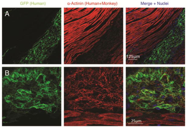
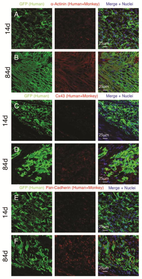
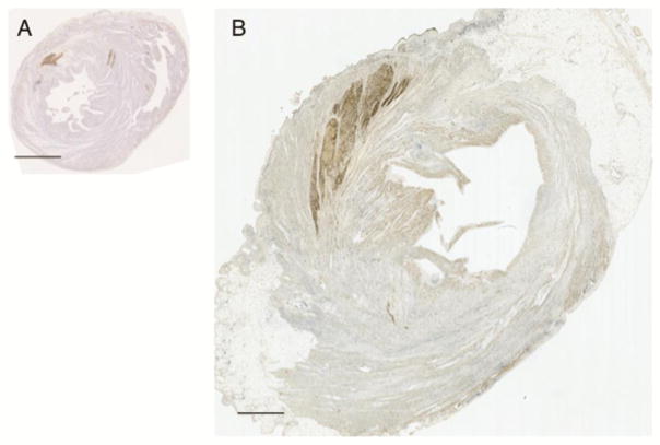
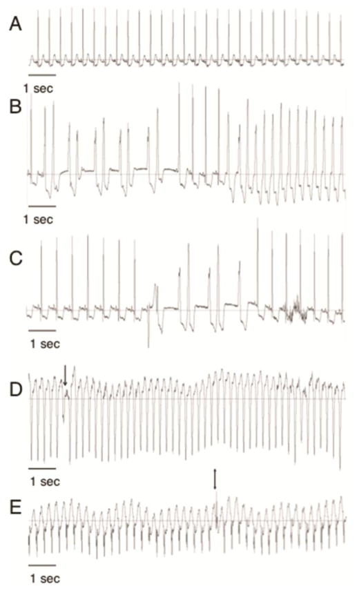
Pluripotent stem cells (PSCs) have indisputable cardiomyogenic potential and therefore have been intensively investigated as a potential cardiac regenerative therapy. Current directed differentiation protocols are able to produce high yields of cardiomyocytes from PSCs and studies in small animal models of cardiovascular disease have proven sustained engraftment and functional efficacy. Therefore, the time is ripe for cardiac regenerative therapies using PSC derivatives to be tested in large animal models that more closely resemble the hearts of humans. In this review, we discuss the results of our recent study using human embryonic stem cell derived cardiomyocytes (hESC-CM) in a non-human primate model of ischemic cardiac injury. Large scale remuscularization, electromechanical coupling and short-term arrhythmias demonstrated by our hESC-CM grafts are discussed in the context of other studies using adult stem cells for cardiac regeneration.


Figures

Figure 1
Figure 1. Human cardiomyocyte grafts within injured monkey hearts
Low power 20× (A) and high power 60× (B) confocal immunofluorescence microscopy of human embryonic stem cell derived cardiomyocytes delivered into the infarcted monkey heart and analysed 14 days after cell delivery. Note the presence of sarcomeric organization and cross-striations present within human cardiomyocytes although much less organized than the host adult monkey cardiomyocytes. GFP= green fluorescent protein.
Figure 2
Figure 2. Human cardiomyocyte grafts display maturation with time after delivery into the host heart
Confocal immunofluorescence of microscopy of human embryonic stem cell derived cardiomyocytes delivered into the infarcted monkey heart. Comparison is made from grafts analysed 14 days (A, C and E) or 84 days (B,D and F) after cell delivery. human cardiomyocytes display increased size and alignment (A–B), increased connexin expression (C–D) and increased cadherin expression (E–F) with time. GFP= green fluorescent protein. Cx43= connexin 43.
Figure 3
Figure 3. Relative size of human cardiomyocyte grafts in the infarcted rat and monkey hearts
Human embryonic stem cell derived cardiomyocyte (hESC-CM) grafts in the infarcted rat (A) and monkey (B) hearts. hESC-CM graft is detected by anti-green fluorescent protein primary antibody with 3,3’-Diaminobenzidine detection of secondary antibody (brown). Scale bar = 2.5mm.
Figure 4
Figure 4. Arrhythmias occur early after engraftment of human cardiomyocytes in the infarcted monkey heart
Monkeys treated with human embryonic stem cell derived cardiomyocytes (hESC-CM) had telemetric electrocardiographic monitoring devices implanted before myocardial infarction. Electrocardiogram traces are shown from the same animal after engraftment of hESC-CM. Sinus rhythm (A) was seen during continuous telemetric recordings before and after myocardial infarction. Early after hESC-CM engraftment non-sustained (duration greater than 3 beats but less than 30 seconds, B–C) and sustained ( duration greater than 30 seconds, D–E) ventricular arrhythmias were observed. Note the different rates. Ventricular tachycardia was defined as a ventricular rhythm with rate of more than 180 beats/min. Slower ventricular rhythms were defined as Accelerated Idioventricular Rhythms. Arrow in (D) shows fusion beat and Double arrow in (E) shows capture beat both of which demonstrate atrioventricular dissociation proving the ventricular origin of the wide-QRS complex rhythm.

References

    1. Askoxylakis V, Thieke C, Pleger ST, Most P, Tanner J, Lindel K, Bischof M. Long-term survival of cancer patients compared to heart failure and stroke: a systematic review. BMC Cancer. 2010;10:105. doi: 10.1186/1471-2407-10-105. - DOI - PMC - PubMed
    1. Assmus B, Schachinger V, Teupe C, Britten M, Lehmann R, Dobert N, Zeiher AM. Transplantation of Progenitor Cells and Regeneration Enhancement in Acute Myocardial Infarction (TOPCARE-AMI) Circulation. 2002;106(24):3009–3017. - PubMed
    1. Balsam LB, Wagers AJ, Christensen JL, Kofidis T, Weissman IL, Robbins RC. Haematopoietic stem cells adopt mature haematopoietic fates in ischaemic myocardium. Nature. 2004;428(6983):668–673. - PubMed
    1. Bel A, Planat-Bernard V, Saito A, Bonnevie L, Bellamy V, Sabbah L, Menasche P. Composite cell sheets: a further step toward safe and effective myocardial regeneration by cardiac progenitors derived from embryonic stem cells. Circulation. 2010;122(11 Suppl):S118–123. doi: 10.1161/CIRCULATIONAHA.109.927293. - DOI - PubMed
    1. Beltrami AP, Barlucchi L, Torella D, Baker M, Limana F, Chimenti S, Anversa P. Adult cardiac stem cells are multipotent and support myocardial regeneration. Cell. 2003;114(6):763–776. - PubMed

Publication types