. 2014 Sep;35(9):1177-87.
doi: 10.1038/aps.2014.58. Epub 2014 Aug 4.

Ursolic acid ameliorates autoimmune arthritis via suppression of Th17 and B cell differentiation

Affiliations

Ursolic acid ameliorates autoimmune arthritis via suppression of Th17 and B cell differentiation

Seung-ye Baek et al. Acta Pharmacol Sin. 2014 Sep.

Abstract

Aim: Ursolic acid (UA) is a pentacyclic triterpenoid found in most plant species, which has been shown anti-inflammatory and anti-oxidative activities. In this study, we examined the effects of UA on collagen-induced arthritis (CIA) in mice, and to identify the mechanisms underlying the effects.

Methods: CIA was induced in mice. Two weeks later, the mice were treated with UA (150 mg/kg, ip, 3 times per week) for 4 weeks. The expression of cytokines and oxidative stress markers in joint tissues was measured with immunohistochemistry. The numbers of CD4+IL-17+, CD4+CD25+Foxp3+ and pSTAT3 cells in spleens were determined using confocal immunostaining or flowcytometric analyses. Serum antibody levels and B cell-associated marker mRNAs were analyzed with ELISAs and qRT-PCR, respectively. CD4+ T cells and CD19+ B cells were purified from mice spleens for in vitro studies.

Results: UA treatment significantly reduced the incidence and severity of CIA-induced arthritis, accompanied by decreased expression of proinflammatory cytokines (TNF-α, IL-1β, IL-6, IL-21 and IL-17) and oxidative stress markers (nitrotyrosine and iNOS) in arthritic joints. In CIA mice, UA treatment significantly decreased the number of Th17 cells, while increased the number of Treg cells in the spleens, which was consistent with decreased expression of pSTAT3, along with IL-17 and RORγt in the splenocytes. In addition, UA treatment significantly reduced the serum CII-specific IgG levels in CIA mice. The inhibitory effects of UA on Th17 cells were confirmed in an in vitro model of Th17 differentiation. Furthermore, UA dose-dependently suppressed the expression of B cell-associated markers Bcl-6, Blimp1 and AID mRNAs in purified CD19+ B cells pretreated with IL-21 or LPS in vitro.

Conclusion: UA treatment significantly ameliorates CIA in mice via suppression of Th17 and differentiation. By targeting pathogenic Th17 cells and autoantibody production, UA may be useful for the treatment of autoimmune arthritis and other Th17-related diseases.


Figures

Figure 1
Figure 1
Ursolic acid (UA) suppresses collagen-induced arthritis (CIA). To assess the influence of UA on CIA, DBA/1J mice were treated with UA (150 mg/kg) or vehicle control three times a week for 4 weeks, beginning 14 d after type II collagen (CII) treatment (n=5 mice per group). (A) Arthritic scores and the incidence of arthritis in CIA-induced DBA/1J mice during the experimental period. (B) Representative histological features of the joints of CIA mice following treatment with either UA or vehicle control. Hematoxylin and eosin (H&E), safranin O, toluidine blue, and TRAP staining results are shown (top). The graph depicts the average histology score and number of TRAP+ cells per joint (bottom). The data are presented as the mean±SD of 6 joints per group. bP<0.05, cP<0.01 relative to the control mice.
Figure 2
Figure 2
UA decreases the CII-specific antibody production in CIA mice. Blood was drawn from the orbital sinuses of UA- and vehicle-treated mice on d 28 and d 42 after first immunization. The concentrations of CII-specific serum IgG and IgG2a were determined by ELISA. The data are presented as the mean±SD. bP<0.05, cP<0.01.
Figure 3
Figure 3
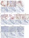
UA decreases the expression of proinflammatory cytokines and oxidative stress-related genes in the joints of CIA mice. Ankle joints (6 per group) of CIA mice treated with either UA or vehicle control were immunostained for (A) IL-1β, IL-6, TNF-α, (B) IL-17, IL-21, nitrotyrosine, and iNOS, or (C) isotype control. Representative data are shown.
Figure 4
Figure 4
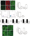
UA reduces STAT3 phosphorylation and decreases the frequency of Th17 cells within the population of CD4+ T cells in CIA mice. (A) Spleens of UA- or vehicle-treated CIA mice were subjected to immunostaining for CD4+IL-17+, CD4+CD25+, and CD4+foxp3+. (B) The frequency CD4+IL-17+ cells among ex vivo total splenocytes was assessed using flow cytometry. (C) The mRNA expression of IL-17, IL-21, and RORγt was determined by real-time RT-PCR in cells obtained from the spleen (Sp) or draining lymph nodes (LN). (D) Spleens of UA- or vehicle-treated CIA mice were subjected to immunostaining of CD4+pSTAT3 Y705+ or CD4+pSTAT3 S727+ cells. The data are presented as the mean±SD of 3 animals per group. bP<0.05, cP<0.01.
Figure 5
Figure 5
UA inhibits Th17 differentiation in vitro. CD4+ T cells were isolated from the spleens of DBA/1J mice and incubated for 3 d under Th17-polarizing conditions (0.5 μg/mL anti-CD3 mAb, 1 μg/mL anti-CD28 mAb, 2 μg/mL anti-IL-4 Ab, 2 μg/mL anti-IFN-γ Ab, 20 ng/mL IL-6, and 2 ng/mL TGF-β) in the presence or absence of UA. (A) UA doses ≤10 μmol/L did not affect cell viability. (B) The frequency of CD4+IL-17+ T cells was measured by flow cytometry. (C) The expression of IL-17, IL-21, RORγt, and Foxp3 was analysed by real-time PCR. The data are presented as the mean±SEM of three independent experiments. bP<0.05, cP<0.01 relative to the control mice.
Figure 6
Figure 6
UA negatively affects B cell function in vitro. (A) Total splenocytes were obtained from DBA/1J mice and stimulated with 1 μg/mL LPS and incubated with various concentrations of UA. After 4 d, the levels of total IgG, IgG1, and IgG2a in the supernatant were determined by ELISAs. (B) CD19+ B cells were isolated from the spleens of DBA/1J mice and pre-treated with either 1 μg/mL LPS or 50 ng/mL IL-21, and then cultured with various concentrations of UA. After 4 d, the mRNA expression of Blimp-1, Bcl-6, and AID was analysed by real-time RT-PCR. bP<0.05, cP<0.01 relative to the controls.

References

    1. McInnes IB, Schett G. The pathogenesis of rheumatoid arthritis. New Engl J Med. 2011;365:2205–19. - PubMed
    1. Egan PJ, van Nieuwenhuijze A, Campbell IK, Wicks IP. Promotion of the local differentiation of murine Th17 cells by synovial macrophages during acute inflammatory arthritis. Arthritis Rheum. 2008;58:3720–9. - PubMed
    1. Mitsdoerffer M, Lee Y, Jäger A, Kim HJ, Korn T, Kolls JK, et al. Proinflammatory T helper type 17 cells are effective B-cell helpers. Proc Natl Acad Sci U S A. 2010;107:14292–7. - PMC - PubMed
    1. Lubberts E, van den Bersselaar L, Oppers-Walgreen B, Schwarzenberger P, Coenen-de Roo CJ, Kolls JK, et al. IL-17 promotes bone erosion in murine collagen-induced arthritis through loss of the receptor activator of NF-kappa B ligand/osteoprotegerin balance. J Immunol. 2003;170:2655–62. - PubMed
    1. Zuniga LA, Jain R, Haines C, Cua DJ. Th17 cell development: from the cradle to the grave. Immunol Rev. 2013;252:78–88. - PubMed

Publication types