

Multiscale optical Ca2+ imaging of tonal organization in mouse auditory cortex
- PMID: 25088366
- PMCID: PMC4242551
- DOI: 10.1016/j.neuron.2014.07.009
Multiscale optical Ca2+ imaging of tonal organization in mouse auditory cortex
Abstract
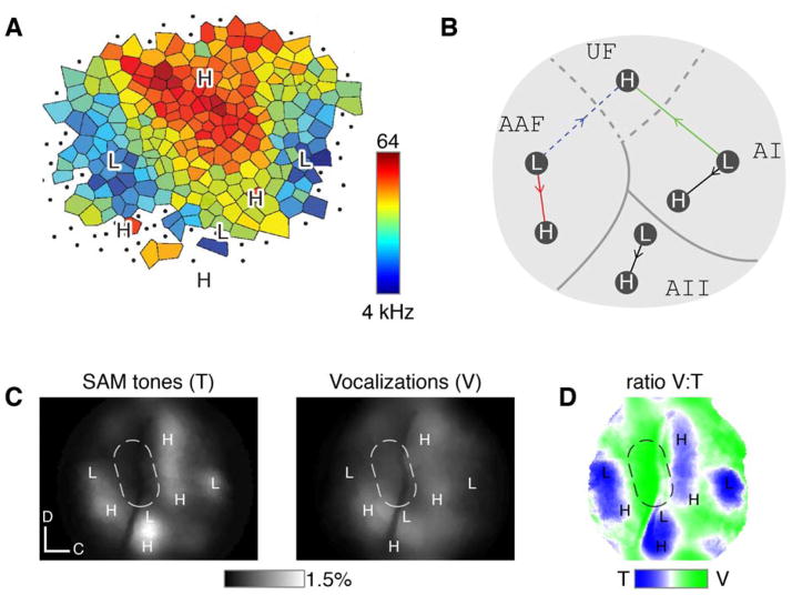
Spatial patterns of functional organization, resolved by microelectrode mapping, comprise a core principle of sensory cortices. In auditory cortex, however, recent two-photon Ca2+ imaging challenges this precept, as the traditional tonotopic arrangement appears weakly organized at the level of individual neurons. To resolve this fundamental ambiguity about the organization of auditory cortex, we developed multiscale optical Ca2+ imaging of unanesthetized GCaMP transgenic mice. Single-neuron activity monitored by two-photon imaging was precisely registered to large-scale cortical maps provided by transcranial widefield imaging. Neurons in the primary field responded well to tones; neighboring neurons were appreciably cotuned, and preferred frequencies adhered tightly to a tonotopic axis. By contrast, nearby secondary-field neurons exhibited heterogeneous tuning. The multiscale imaging approach also readily localized vocalization regions and neurons. Altogether, these findings cohere electrode and two-photon perspectives, resolve new features of auditory cortex, and offer a promising approach generalizable to any cortical area.
Copyright © 2014 Elsevier Inc. All rights reserved.
Figures


Comment in
-
Measuring the functional organization of the neocortex at large and small scales.Neuron. 2014 Aug 20;83(4):756-8. doi: 10.1016/j.neuron.2014.08.008. Neuron. 2014. PMID: 25144871
References
Publication types
MeSH terms
Substances
Grants and funding
LinkOut - more resources
Full Text Sources
Other Literature Sources
Molecular Biology Databases
Miscellaneous
