Review
. 2014 Aug;40(8):1263-72.
doi: 10.1016/j.jcrs.2014.06.020.

Aqueous outflow: segmental and distal flow

Affiliations
Review

Aqueous outflow: segmental and distal flow

Swarup S Swaminathan et al. J Cataract Refract Surg. 2014 Aug.

Abstract

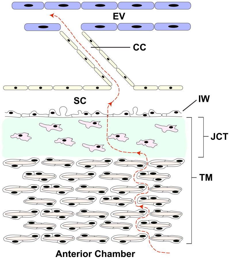
The elevated intraocular pressure (IOP) of primary open-angle glaucoma is caused by impaired outflow of aqueous humor through the trabecular meshwork. Within the juxtacanalicular region, alterations of both extracellular matrix homeostasis and the cellular tone of trabecular meshwork endothelial and the inner wall of Schlemm canal cells affect outflow. Newer pharmacologic agents that target trabecular meshwork and Schlemm canal cell cytoskeleton lower IOP. Aqueous drainage occurs nonhomogenously with greater flow going through certain portions of the TM and less going through other portions-a concept known as segmental flow, which is theoretically the result of outflow being dependent on the presence of discrete pores within Schlemm canal. The limited long-term success of trabecular meshwork bypass surgeries implicates the potential impact of resistance in Schlemm canal itself and collector channels. Additionally, others have observed that outflow occurs preferentially near collector channels. These distal structures may be more important to aqueous outflow than previously believed.

Financial disclosure: Dr. Rhee is a consultant to Aerie Pharmaceuticals, Alcon Laboratories, Inc., Allegan, Inc., Aquesys, Inc., Glaukos Corp., Ivantis, Inc., Johnson & Johnson, Merck Sharp & Dohme Corp. and Santen, Inc., and has received research funding from Alcon Laboratories, Inc., Merck Sharp & Dohme Corp., and Ivantis, Inc. No other author has a financial or proprietary interest in any material or method mentioned.


Figures

Figure 1
Figure 1
Schematic illustrating the major components of the conventional outflow pathway. Aqueous humor (red dashed line) flows through the initial portion of the trabecular meshwork (TM), juxtacanalicular connective tissue region, inner wall of Schlemm canal (IW), Schlemm canal (SC), collector channel (CC), and finally reaches the episcleral vein (EV). Multiple TM cells encase the trabecular beams (tan) within the TM. The juxtacanalicular connective tissue is composed of sparse cells (pink) and substantial ECM (green).
Figure 2
Figure 2
Impact of ROCK inhibitor on trabecular meshwork cell morphology and shape (cells stained with phalloidin). Actin is prominent in (A) normal cells, but is significantly decreased in (B) cells treated with the ROCK inhibitor Y-27632. (Inoue T, Tanihara H. Rho-associated kinase inhibitors: a novel glaucoma therapy. Prog Retin Eye Res 2013; 37:1–12, reprinted with permission from Elsevier.)
Figure 3
Figure 3
The effect of SPARC overexpression on collagen IV expression within the trabecular meshwork in perfused human cadaveric eyes. The SPARC overexpression is confirmed (red), and the concentration of collagen IV is significantly increased with SPARC overexpression (green) (bar = 30μm).
Figure 4
Figure 4
Electron microscopy of the juxtacanalicular connective tissue. This portion of the trabecular meshwork is composed of sparse cells (C) with abundant, amorphous extracellular matrix (*). The inner wall of Schlemm canal (arrow) and Schlemm canal (SC) are labeled.
Figure 5
Figure 5
Electron microscopy demonstrating pores (arrows) within the inner wall endothelium of Schlemm canal. (Reprinted with permission. Allingham RR, de Kater AW, Ethier CR, Anderson PJ, Hertzmark E, Epstein DL. The relationship between pore density and outflow facility in human eyes. Invest Ophthalmol Vis Sci 1992; 33:1661– 1669. Available at: http://www.iovs.org/content/33/5/1661.full.pdf. Accessed June 16, 2014
Figure 6
Figure 6
Schematic of funneling theory, whereby outflow through the trabecular meshwork occurs specifically in areas in which pores in the inner wall of Schlemm canal are present. Reprinted with permission from Investigative Ophthalmology and Visual Sciences.
Figure 7
Figure 7
Illustration of segmental flow in the mouse eye using fluorescent microbeads. A: Segmental flow in the wild-type mouse eye. Microbeads are only present in certain parts of the trabecular meshwork. B: Outflow is more uniform in the SPARC knockout mouse due to the lack of this matricellular protein.
Figure 8
Figure 8
Effect of ROCK inhibitor on the association between juxtacanalicular connective tissue and inner wall of Schlemm canal of monkey eyes. A: Normal, untreated eye with Schlemm canal (SC) and trabecular meshwork (TM) labeled. It is difficult to distinguish the inner wall from the TM. B: Eye treated with a ROCK inhibitor. There is a clear separation of the inner wall, with a ballooning effect of the juxtacanalicular connective tissue region. Reprinted with permission from Experimental Eye Research.
Figure 9
Figure 9
Affinity of fluorescent microbeads for trabecular meshwork near collector channels. A: Fluorescent microbeads preferentially collect within the trabecular meshwork (TM) near collector channels (CC). B: Increase in microbead concentration in this area after treatment with a ROCK inhibitor. Reprinted with permission from Experimental Eye Research.
Figure 10
Figure 10
Light microscopy of trabecular meshwork of bovine eyes perfused at various pressures. A: At 7mm Hg, the aqueous plexus (AP) and collector channel (CC) are well organized. B, C, D: At progressively higher perfusion pressures, herniation of the trabecular meshwork into the AP and CC is observed. Reprinted with permission from Investigative Ophthalmology and Visual Sciences.

References

    1. Weinreb RN, Khaw PT. Primary open-angle glaucoma. Lancet. 2004;363:1711–1720. - PubMed
    1. Quigley HA. Number of people with glaucoma worldwide. [Accessed June 13, 2014];Br J Ophthalmol. 1996 80:389–393. Available at: http://www.ncbi.nlm.nih.gov/pmc/articles/PMC505485/pdf/brjopthal00005-00.... - PMC - PubMed
    1. Rohen JW. Why is intraocular pressure elevated in chronic simple glaucoma? Anatomical considerations Ophthalmology. 1983;90:758–765. - PubMed
    1. Bill A, Phillips CI. Uveoscleral drainage of aqueous humour in human eyes. Exp Eye Res. 1971;12:275–281. - PubMed
    1. Overby DR, Stamer WD, Johnson M. The changing paradigm of outflow resistance generation: towards synergistic models of the JCT and inner wall endothelium. Exp Eye Res. 2009;88:656–670. - PMC - PubMed

Other Cited Material

    1. Vranka J, Keller KE, Acott T. Extracellular matrix gene expression profiling of high and low flow areas of human trabecular meshwork. [Accessed June 13, 2014];IOVS. 2013 54 ARVO E-Abstract 3566. Available at: http://abstracts.iovs.org//cgi/content/abstract/54/6/3566?sid=c784a366-6....

MeSH terms

Substances