. 2014 Aug 21;41(2):283-95.
doi: 10.1016/j.immuni.2014.06.016. Epub 2014 Jul 31.

MHCII-mediated dialog between group 2 innate lymphoid cells and CD4(+) T cells potentiates type 2 immunity and promotes parasitic helminth expulsion

Affiliations

MHCII-mediated dialog between group 2 innate lymphoid cells and CD4(+) T cells potentiates type 2 immunity and promotes parasitic helminth expulsion

Christopher J Oliphant et al. Immunity. .

Abstract

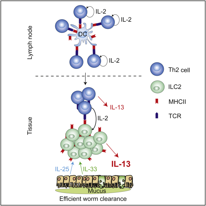
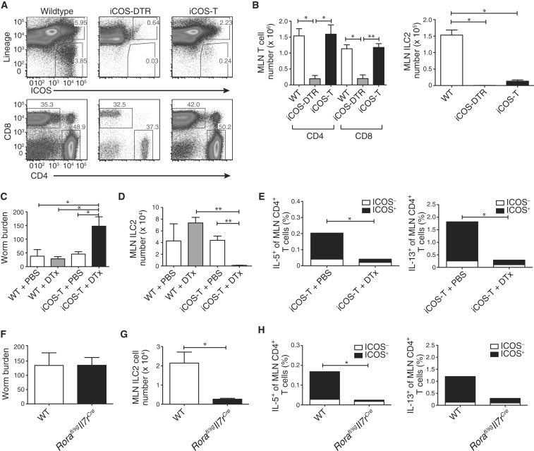
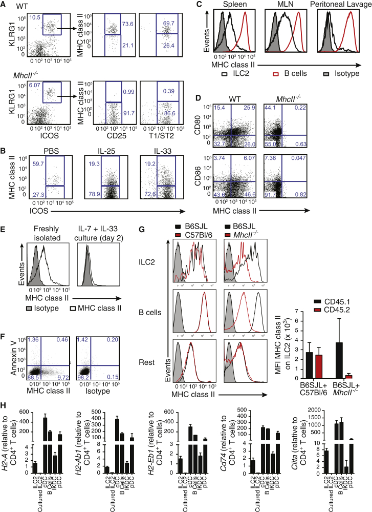
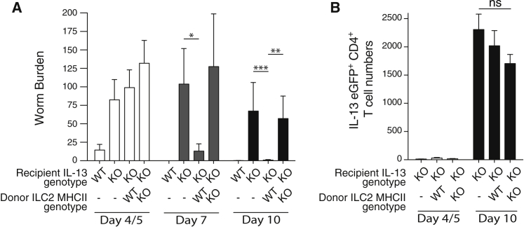
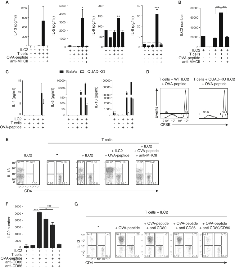
Group 2 innate lymphoid cells (ILC2s) release interleukin-13 (IL-13) during protective immunity to helminth infection and detrimentally during allergy and asthma. Using two mouse models to deplete ILC2s in vivo, we demonstrate that T helper 2 (Th2) cell responses are impaired in the absence of ILC2s. We show that MHCII-expressing ILC2s interact with antigen-specific T cells to instigate a dialog in which IL-2 production from T cells promotes ILC2 proliferation and IL-13 production. Deletion of MHCII renders IL-13-expressing ILC2s incapable of efficiently inducing Nippostrongylus brasiliensis expulsion. Thus, during transition to adaptive T cell-mediated immunity, the ILC2 and T cell crosstalk contributes to their mutual maintenance, expansion and cytokine production. This interaction appears to augment dendritic-cell-induced T cell activation and identifies a previously unappreciated pathway in the regulation of type-2 immunity.


Figures

None
Graphical abstract
Figure 1
Figure 1
ILC2 Ablation Leads to Impaired Th2 Responses to N. brasiliensis (A) ILC2 ablation. Representative flow cytometric analysis of MLN from WT, iCOS-DTR (without Cd4Cre), or iCOS-T mice (with Cd4Cre), treated with three daily doses of DTx (25 ng/g bodyweight) and IL-33. (B) Numbers of T cells and ILC2s in MLN of mice, treated daily with 3 doses of DTx and IL-33. (C) Worm burden in mice at 5 days postinfection (d.p.i). iCOS-T mice treated daily with DTx (15 ng/g bodyweight, i.p.). (D) ILC2s in mice 5 d.p.i. (E) CD4+ T cells producing IL-5 and IL-13. Statistical analysis performed between CD4+ICOS+ T cell populations (black bars). (F) Worm burden in Rorafl/sgIl7rCre mice 5 d.p.i. (G) ILC2 numbers in Rorafl/sgIl7rCre mice 5 d.p.i. (H) CD4+ T cells producing IL-5 and IL-13. Statistical analysis performed between CD4+ICOS+ T cell populations (black bars). Data are representative of at least two independent experiments with three (A and B) or five (C–H) mice per group and bar graphs represent mean ± SEM. p < 0.05 and ∗∗ < 0.001. See also Figures S1 and S2.
Figure 2
Figure 2
MHCII Is Expressed by ILC2s (A) Representative flow cytometry of MHCII on ILC2s from the MLN of naive mice. Plots are gated on lin cells. (B) MHCII expression by ILC2s from the MLN of mice treated i.p. as indicated. Plots are gated on lin cells. (C) MHCII expression on IL-33-elicited ILC2s and B cells from the indicated tissues. (D) CD80 and CD86 expression on LinICOS+ MLN cells from IL-33-treated mice. (E) MHCII expression on IL-25-elicited ILC2s cultured as indicated. (F) Annexin V and MHCII expression on IL-33-elicited lymph node ILC2s cultured for 48 hr. (G) Chimeric mice were generated by mixing B6SJL (CD45.1+) bone marrow with either C57Bl/6 or MhcII−/− bone marrow (CD45.2+) in a 1:1 ratio. MHCII expression was determined following three doses of IL-33 (i.p.). Graph shows mean fluorescence intensity (MFI) of MHCII expression by ILC2s. (H) Quantitative RT-PCR for MHCII (H2-Aa, H2-Ab1, or H2-Eb1), Cd74 and Ciita gene expression from the indicated populations. ILC2s are from the MLN of IL-33-treated mice. Data are representative of at least two independent experiments with three mice per group. Bar graphs represent mean ± SEM. See also Figure S3.
Figure 3
Figure 3
MHCII-Deficient ILC2s Fail to Rescue N. brasiliensis Expulsion in IL-13-Deficient Hosts (A) Worm burden in IL-13-deficient hosts following transfer of ILC2s from IL-25-treated WT or MhcII−/− donors. Data are pooled from four independent experiments with five or six mice in each group per experiment. p < 0.05, ∗∗p < 0.01, and ∗∗∗p < 0.001. (B) Number of Il13egfp/egfp CD4+ T cells in MLN following infection. Data are representative of three independent experiments with five to six mice per group. Bar graphs represent mean ± SEM. See also Figure S4.
Figure 4
Figure 4
Antigen-Dependent Activation of T Cells by ILC2s Requires MHCII (A) Cell trace violet (CTV)-labeled OTIITg CD4+ T cells were cultured in a 1:1 ratio with IL-33-elicited ILC2s, pulsed with or without OVA-peptide. (B) Antigen-dependent OTIITg CD4+ T cell proliferation induced by the indicated populations. (C) CFSE-labeled OTIITg CD4+ T cell proliferation, as indicated. (D) CTV-labeled OTIITg CD4+ T cell proliferation, as indicated. (E) OTIITg CD4+ T cell numbers in cocultures, as indicated. (F and G) The number of activated OTIITg CD4+ T cells (CD44+CD69+ or CD44+CD62L) following coculture with antigen-pulsed ILC2s. (H) CTV-labeled OTIITg CD4+ T cell proliferation, as indicated. Data are representative of three independent experiments with two or three mice per group. (I) Detection of processed and presented Eα peptide on ILC2s. Data are representative of two independent experiments. ILC2s were elicited using IL-33 and isolated from mesenteric, para-aortic, and inguinal lymph nodes. Bar graphs represent mean ± SEM. See also Figure S5.
Figure 5
Figure 5
ILC2s Enter Antigen-Dependent Dialog with CD4+ T Cells (A) Cytokines in supernatants of cocultures containing peptide-pulsed ILC2s and OTIITg CD4+ T cells. Dotted line indicates limit of detection for each assay. (B) ILC2 number following coculture with OTIITg CD4+ T cells, as indicated. (C) Cytokine production in supernatants of WT (BALB/c) or QUAD-KO ILC2s and DO11.10Tg T cell cocultures, as indicated. (D) CFSE-labeled DO11.10Tg T cell proliferation following coculture with QUAD-KO ILC2s, as indicated. (E) Intracellular IL-13 staining following ILC2:OTIITg CD4+ T cell coculture, as indicated. (F) ILC2 number following coculture with OTIITg CD4+ T cells, as indicated. (G) Intracellular IL-13 staining following ILC2:OTIITg CD4+ T cell coculture, as indicated. Data are representative of three independent experiments with two or three mice per group. MLN ILC2s were elicited using IL-33 (A, B, E–G) or IL-25 (C and D). Bar graphs represent mean ± SEM. See also Figure S6.
Figure 6
Figure 6
IL-2 Is Required for the Antigen-Dependent Dialog between ILC2s and T Cells (A and B) Number of IL-13-expressing ILC2s (A) and T cells (B) from ILC2:OTIITg CD4+ T cell cocultures in the presence of IL-4-blocking antibodies. (C) Surface CD25 and intracellular IL-13 expression by ILC2s stimulated with IL-2 for 72 hr. (D) Intracellular IL-2 staining of CTV-labeled OTIITg CD4+ T cells following coculture with OVA-peptide-loaded WT ILC2s. (E) Number of IL-2-expressing T cells from ILC2:OTIITg CD4+ T cell cocultures in the presence of MHCII-blocking antibodies. (F) IL-13 concentration in supernatants from ILC2:OTIITg CD4+ T cell cocultures in the presence of blocking anti-IL-2 antibodies. (G and H) Number of cytokine expressing ILC2s (G) and OTIITg CD4+ T cells (H) in the presence of IL-2 blocking antibodies. (I) Worm burdens and (J) ILC2s in Rag2−/− mice 10 d.p.i. with N. brasiliensis and treated as indicated. Lymph node ILC2s were elicited with IL-33. Bar graphs represent mean ± SEM. Data are representative of three independent experiments with two or three mice per group (A–H) or from a single experiment with five or six mice per group (I and J). Bar graphs represent mean ± SEM.

Comment in

References

    1. Barlow J.L., Bellosi A., Hardman C.S., Drynan L.F., Wong S.H., Cruickshank J.P., McKenzie A.N. Innate IL-13-producing nuocytes arise during allergic lung inflammation and contribute to airways hyperreactivity. J. Allergy Clin. Immunol. 2012;129:191–198. e191-194. - PubMed
    1. Barnden M.J., Allison J., Heath W.R., Carbone F.R. Defective TCR expression in transgenic mice constructed using cDNA-based alpha- and beta-chain genes under the control of heterologous regulatory elements. Immunol. Cell Biol. 1998;76:34–40. - PubMed
    1. Bartemes K.R., Iijima K., Kobayashi T., Kephart G.M., McKenzie A.N., Kita H. IL-33-responsive lineage- CD25+ CD44(hi) lymphoid cells mediate innate type 2 immunity and allergic inflammation in the lungs. J. Immunol. 2012;188:1503–1513. - PMC - PubMed
    1. Chan C.W., Crafton E., Fan H.N., Flook J., Yoshimura K., Skarica M., Brockstedt D., Dubensky T.W., Stins M.F., Lanier L.L. Interferon-producing killer dendritic cells provide a link between innate and adaptive immunity. Nat. Med. 2006;12:207–213. - PubMed
    1. Chang Y.J., Kim H.Y., Albacker L.A., Baumgarth N., McKenzie A.N., Smith D.E., Dekruyff R.H., Umetsu D.T. Innate lymphoid cells mediate influenza-induced airway hyper-reactivity independently of adaptive immunity. Nat. Immunol. 2011;12:631–638. - PMC - PubMed

Publication types

MeSH terms