. 2014 Oct:144 Pt B:402-9.
doi: 10.1016/j.jsbmb.2014.07.011. Epub 2014 Jul 30.

The vitamin D system is deregulated in pancreatic diseases

Affiliations

The vitamin D system is deregulated in pancreatic diseases

Doris Hummel et al. J Steroid Biochem Mol Biol. 2014 Oct.

Abstract

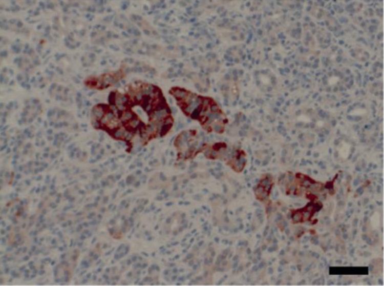
The vitamin D system is deregulated during development and progression of several cancer types. Data on the expression of the vitamin D system in the diseased pancreas are missing. The aim of this study was to investigate the expression of the vitamin D receptor (VDR), 1,25-dihydroxyvitamin D3 24-hydroxylase (CYP24A1), and the calcium-sensing receptor (CaSR), a vitamin D target gene, in the different regions of the pancreas in patients with chronic pancreatitis (n=6) and pancreatic ductal adenocarcinomas (PDAC) (n=17). We analyzed the expression of these genes at mRNA and protein level with quantitative real-time RT-PCR and immunostaining. mRNA expression of CYP24A1 and VDR was significantly increased in tumors compared with the adjacent non-tumorous tissue (p<0.01), while CaSR mRNA expression decreased. Both the VDR and the CaSR protein were highly expressed in the endocrine compared with the exocrine pancreas. In CP the CYP24A1 expression was highest in the endocrine pancreas, while in PDACs in the transformed ducts. In the PDAC patients CYP24A1 expression in the islets was significantly lower than in CP patients. Our data suggest that during ductal adenocarcinoma development the vitamin D system in the pancreas becomes deregulated on two levels: in the islets CYP24A1 expression decreases weakening the negative feedback regulation of the vitamin D-dependent insulin synthesis/secretion. In the transformed ducts CYP24A1 expression increases, impairing the antiproliferative effect of vitamin D in these cells.

Keywords: CYP24A1; CaSR; Chronic pancreatitis; Pancreatic cancer; Pancreatic ductal adenocarcinoma; VDR; Vitamin D.


Figures

Fig. 1
Fig. 1
mRNA expression of the VDR and CYP24A1 is significantly increased and CaSR expression decreases in PDAC compared with adjacent non-tumorous (NT) parts of the pancreas. (A) mRNA expression (n = 11) was assessed by qRT-PCR and set relative to calibrator. (B) Data originate from a microarray analysis of mRNA from patients suffering from PDAC . Median, interquartile range and whiskers according to Tukey are shown. Statistics were calculated using paired t-test on log2-transformed data. Among the values for CYP24A1 expression we identified one outlier in each dataset. (**p < 0.01, ***p < 0.001).
Fig. 2
Fig. 2
Protein expression of VDR in CP and PDAC. VDR is highly expressed in endocrine islets compared with other parts of the pancreas. (A) IHC of an endocrine islet in a region of chronic pancreatitis is shown. Black bar equals 50 μm. (B) Protein expression was determined using IHC, percentage of VDR positive cells is shown. Median, interquartile range and whiskers according to Tukey are shown. Wilcoxon signed ranks test was applied to compare protein expression in patients with CP and PDAC, and Mann–Whitney U-test to compare between disease groups. Both tests were followed by Bonferroni–Holm correction for multiple comparisons. Asterisks above boxes indicate statistically significant difference from stroma (*< 0.05).
Fig. 3
Fig. 3
Expression of CYP24A1 in pancreatic ductal adenocarcinoma compared with non-tumorous regions of the same patient. (A) IHC of CYP24A1 in a patient with PDAC is shown. Black bar equals 50 μM. (B) Protein expression of the CYP24A1 is increased in ductal adenocarcinoma compared with other regions of pancreatic tissue. Protein expression was determined using IHC, percentage of CYP24A1 positive cells is shown. Median, interquartile range and whiskers according to Tukey are shown. Wilcoxon signed ranks test was applied to compare protein expression in patients with CP and PDAC, and Mann–Whitney U-test to compare between disease groups. Both tests were followed by Bonferroni–Holm correction for multiple comparisons. Asterisks above boxes indicate statistically significant difference from stroma (*p < 0.05). Hashtags indicate statistically significant difference between patients with CP and PDAC (#< 0.05, ##< 0.01). (For interpretation of the references to color in this figure legend, the reader is referred to the web version of this article.)
Fig. 4
Fig. 4
Expression of Ki67 in chronic pancreatitis and pancreatic ductal adenocarcinoma. CP patients have low expression of the proliferative marker Ki67. In PDAC patients Ki67 expression is increased in tumor compared with non-tumorous regions of the same patient. Immunostaining of Ki67 (brown) in patient with CP (left) and PDAC (right) is shown. (For interpretation of the references to color in this figure legend, the reader is referred to the web version of this article.)
Fig. 5
Fig. 5
CaSR expression is high in endocrine islets compared with other parts of the pancreas. IHC of an endocrine islet with surrounding acinar and duct cells is shown. Black bar equals 50 μm.
Fig. 6
Fig. 6
VDR and the CaSR are expressed in the endocrine cells of the pancreas. (A) Immunofluorescence staining of the endocrine marker synaptophysin (SYN, green) and the VDR (red) and the merged picture. (B) Immunofluorescence staining of the CaSR (white) and the VDR (red) and the merged picture. White bar equals 50 μM.

References

    1. Hidalgo M. Pancreatic cancer. New Engl. J. Med. 2010;362:1605–1617. - PubMed
    1. Rosen C.J., Adams J.S., Bikle D.D., Black D.M., Demay M.B., Manson J.E., Murad M.H., Kovacs C.S. The nonskeletal effects of vitamin D: an endocrine society scientific statement. Endocr. Rev. 2012;33:456–492. - PMC - PubMed
    1. Norman A.W. From vitamin D to hormone D: fundamentals of the vitamin D endocrine system essential for good health. Am. J. Clin. Nutr. 2008;88 491S–499S. - PubMed
    1. Townsend K., Evans K.N., Campbell M.J., Colston K.W., Adams J.S., Hewison M. Biological actions of extra-renal 25-hydroxyvitamin D-1alpha-hydroxylase and implications for chemoprevention and treatment. J. Steroid Biochem. Mol. Biol. 2005;97:103–109. - PubMed
    1. Pike J.W., Meyer M.B. The vitamin D receptor: new paradigms for the regulation of gene expression by 1,25-dihydroxyvitamin D3. Rheum. Dis. Clin. North Am. 2012;38:13–27. - PMC - PubMed

Publication types