. 2014 Aug 6:12:218.
doi: 10.1186/s12967-014-0218-x.

A microRNA profile of human CD8(+) regulatory T cells and characterization of the effects of microRNAs on Treg cell-associated genes

Affiliations

A microRNA profile of human CD8(+) regulatory T cells and characterization of the effects of microRNAs on Treg cell-associated genes

Fadi Jebbawi et al. J Transl Med. .

Abstract

Background: Recently, regulatory T (Treg) cells have gained interest in the fields of immunopathology, transplantation and oncoimmunology. Here, we investigated the microRNA expression profile of human natural CD8(+)CD25(+) Treg cells and the impact of microRNAs on molecules associated with immune regulation.

Methods: We purified human natural CD8(+) Treg cells and assessed the expression of FOXP3 and CTLA-4 by flow cytometry. We have also tested the ex vivo suppressive capacity of these cells in mixed leukocyte reactions. Using TaqMan low-density arrays and microRNA qPCR for validation, we could identify a microRNA 'signature' for CD8(+)CD25(+)FOXP3(+)CTLA-4(+) natural Treg cells. We used the 'TargetScan' and 'miRBase' bioinformatics programs to identify potential target sites for these microRNAs in the 3'-UTR of important Treg cell-associated genes.

Results: The human CD8(+)CD25(+) natural Treg cell microRNA signature includes 10 differentially expressed microRNAs. We demonstrated an impact of this signature on Treg cell biology by showing specific regulation of FOXP3, CTLA-4 and GARP gene expression by microRNA using site-directed mutagenesis and a dual-luciferase reporter assay. Furthermore, we used microRNA transduction experiments to demonstrate that these microRNAs impacted their target genes in human primary Treg cells ex vivo.

Conclusions: We are examining the biological relevance of this 'signature' by studying its impact on other important Treg cell-associated genes. These efforts could result in a better understanding of the regulation of Treg cell function and might reveal new targets for immunotherapy in immune disorders and cancer.


Figures

Figure 1
Figure 1
Purified human CD8 + CD25 + natural Tregs express FOXP3 and CTLA-4. Separated cell fractions, CD8+CD25+ and CD8+CD25 T cells, were analyzed by multicolor FACS using the following antibodies: anti-CD3 PerCP, anti-CD8-APC (BD biosciences) and anti-CD25-PE (Miltenyi Biotec) to assess purity for each isolation. Intracellular FOXP3 and cell-surface CTLA-4 were assessed using human anti-FOXP3-PE detection kit and anti-CTLA-4-PE (BD biosciences). Flow cytometry was performed using a FACSCalibur applying CellQuest software (BD Biosciences). CD3 and CD8 purity was above 97% among isolated CD8+ T cells. CD8+CD25+ nTregs express FOXP3 and CTLA-4 when compared to CD8+CD25 T cells.
Figure 2
Figure 2
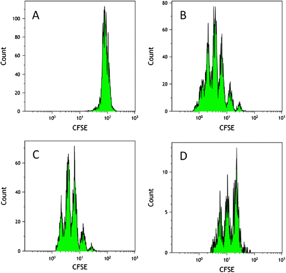
Functional assessment of CD8 + CD25 + nTreg cell suppressive capacity toward proliferation of allogeneically activated CFSE-labeled T lymphocytes. CFSE dilution analysis shows CD8+CD25+ nTreg-mediated suppression of allogeneic T cell proliferation in 5 days mixed leukocyte reaction (MLR) compared to control MLR and compared to MLR with CD8+CD25 non-Tregs. CFSE dilution histograms are shown for 1:1 suppressor/responder cell ratio. Shown is one representative experiment out of four independent experiments performed. (A) CFSE-labeled allogeneic T cells alone at d0 before MLR. (B) Proliferation of CFSE-labeled allogeneic T cells after 5 days of allo-MLR (control MLR). (C) Allo-MLR in presence of CD8+CD25 non-Treg cells, at day 5. (D) Allo-MLR in presence of CD8+CD25+ nTregs, at day 5.
Figure 3
Figure 3
Differential expression of miR-24, −335, −155, −31, −210, −449, −509, −214, −205 and −9 between CD8 + CD25 + nTregs and CD8 + CD25 T cells. Data obtained by qRT-PCR amplification of miRs are plotted. p values for each miRNA relative expression are presented. Boxes represent SE; and Error bars SD Pooled data from five independent experiments are shown. (*p < 0.05; **p < 0.01 CD8+CD25+ nTregs versus CD8+CD25 T cells (Student's t test).
Figure 4
Figure 4
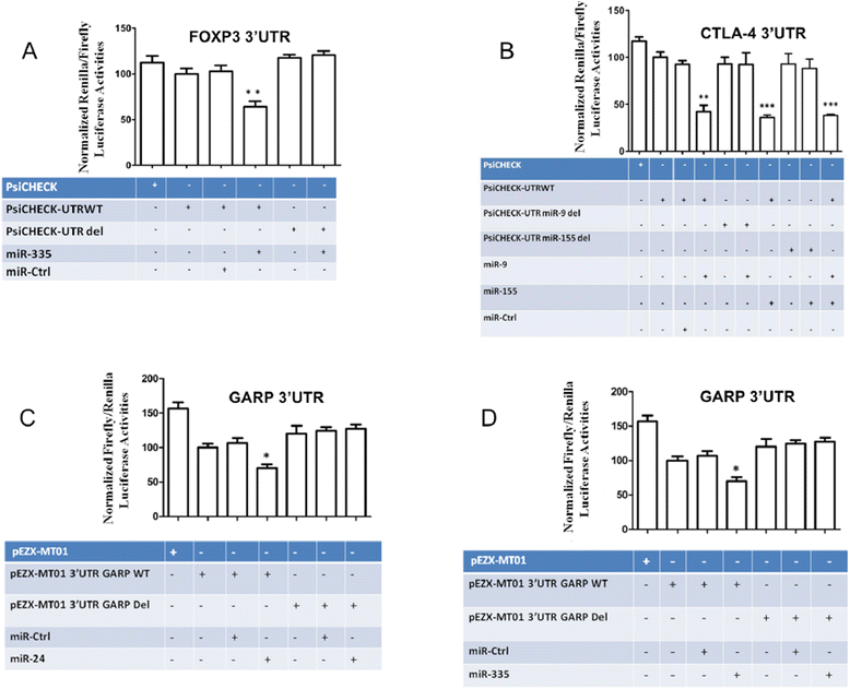
MicroRNA specific activities. (A) MiR-335 negatively regulates luciferase expression in a plasmid coupling its coding sequence with FOXP3 3′UTR. Renilla luciferase reporter assays with constructs holding FOXP3 3′-UTR sequences from the indicated genes were co-transfected into HEK293T cells along with a firefly luciferase transfection control plasmid either alone or together with miR-335. (B) MiR-9 and miR-155 negatively regulate luciferase expression in a plasmid coupling its coding sequence with CTLA-4 3′UTR. Renilla luciferase reporter assays with constructs holding CTLA-4 3′-UTR sequences, wild type or miR-site deleted, were co-transfected into HEK293T cells along with a firefly luciferase transfection control plasmid either alone or together with miR-9, −155. MiR-24 (C) and miR-335 (D) specifically targets GARP 3′UTR and negatively regulate luciferase reporter expression. Renilla and firefly luciferase reporter assays with constructs holding GARP 3′-UTR sequences, wild type or miR-site deleted, were co-transfected into HEK293T cells along with miR-24, −335. Relative luciferase values normalized to transfections without miRNA are shown. Data represent mean ± SD (error bars) of three independent experiments, each performed in triplicate. (*p < 0.05; **, p < 0.01, Student's t test).
Figure 5
Figure 5
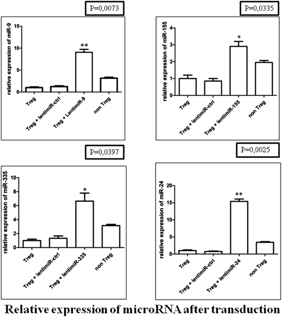
Differential expression of miR-24, −335, −155 and −9 in CD8 + CD25 + natural Treg cells after transduction by lenti-miR-24, −335, −155 and −9. Data obtained by qRT-PCR amplification of miRs are plotted for CD8+CD25+nTregs versus non-transduced CD8+CD25+ nTregs. p values for each miRNA relative expression are presented. Boxes represent SE; and Error bars SD. Pooled data from five independent experiments are shown. (*p < 0.05; **p < 0.01 Student's t test).
Figure 6
Figure 6
MicroRNA specific activities. (A) In primary human Treg cells, miR-335 specifically regulates FOXP3 expression. Relative miR-335 and FOXP3 expression in CD8+CD25+ Treg cells transduced by lenti-miR-335 compared with CD8+CD25+ Treg cells transduced by lenti-miR-Ctrl, as determined by relative qRT-PCR. (B) In primary human Treg cells, miR-9 and −155 specifically regulate CTLA-4 expression. Relative miR-9, miR-155 and CTLA-4 expression in CD8+CD25+ Treg cells transduced by lenti-miR-9 or lenti-miR-155 compared with CD8+CD25+ Treg cells transduced by lenti-miR-Ctrl, as determined by relative qRT-PCR. (C) In primary human Treg cells, miR-24 and −335 specifically regulate GARP expression. Relative miR-24 and GARP expression in CD8+CD25+ Treg cells transduced by lenti-miR-24 compared with CD8+CD25+ Treg cells transduced by lenti-miR-Ctrl, as determined by relative qRT-PCR. Relative miR-335 and GARP expression in CD8+CD25+ Treg cells transduced by lenti-miR-335 compared with CD8+CD25+ Treg cells transduced by lenti-miR-Ctrl. as determined by relative qRT-PCR. CD8+CD25 T cells were considered to be non-Tregs; *p < 0.05, **p < 0.01, ***p < 0.001 (as determined by Student's t-test).

References

    1. Dinesh RK, Skaggs BJ, La Cava A, Hahn BH, Singh RP. CD8+ Tregs in lupus, autoimmunity, and beyond. Autoimmun Rev. 2010;9(8):560–568. doi: 10.1016/j.autrev.2010.03.006. - DOI - PMC - PubMed
    1. Daniele N, Scerpa MC, Landi F, Caniglia M, Miele MJ, Locatelli F, Isacchi G, Zinno F. T(reg) cells: collection, processing, storage and clinical use. Pathol Res Pract. 2011;207(4):209–215. doi: 10.1016/j.prp.2011.02.003. - DOI - PubMed
    1. Huseby ES, Liggitt D, Brabb T, Schnabel B, Ohlen C, Goverman J. A pathogenic role for myelin-specific CD8(+) T cells in a model for multiple sclerosis. J Exp Med. 2001;194(5):669–676. doi: 10.1084/jem.194.5.669. - DOI - PMC - PubMed
    1. Liblau RS, Wong FS, Mars LT, Santamaria P. Autoreactive CD8 T cells in organ-specific autoimmunity: emerging targets for therapeutic intervention. Immunity. 2002;17(1):1–6. doi: 10.1016/S1074-7613(02)00338-2. - DOI - PubMed
    1. Miller A, Lider O, Roberts AB, Sporn MB, Weiner HL. Suppressor T cells generated by oral tolerization to myelin basic protein suppress both in vitro and in vivo immune responses by the release of transforming growth factor beta after antigen-specific triggering. Proc Natl Acad Sci U S A. 1992;89(1):421–425. doi: 10.1073/pnas.89.1.421. - DOI - PMC - PubMed

Publication types

MeSH terms