. 2015 Jan;25(1):8-20.
doi: 10.1093/glycob/cwu080. Epub 2014 Aug 4.

RNAi screening of human glycogene orthologs in the nematode Caenorhabditis elegans and the construction of the C. elegans glycogene database

Affiliations

RNAi screening of human glycogene orthologs in the nematode Caenorhabditis elegans and the construction of the C. elegans glycogene database

Sayaka Akiyoshi et al. Glycobiology. 2015 Jan.

Abstract

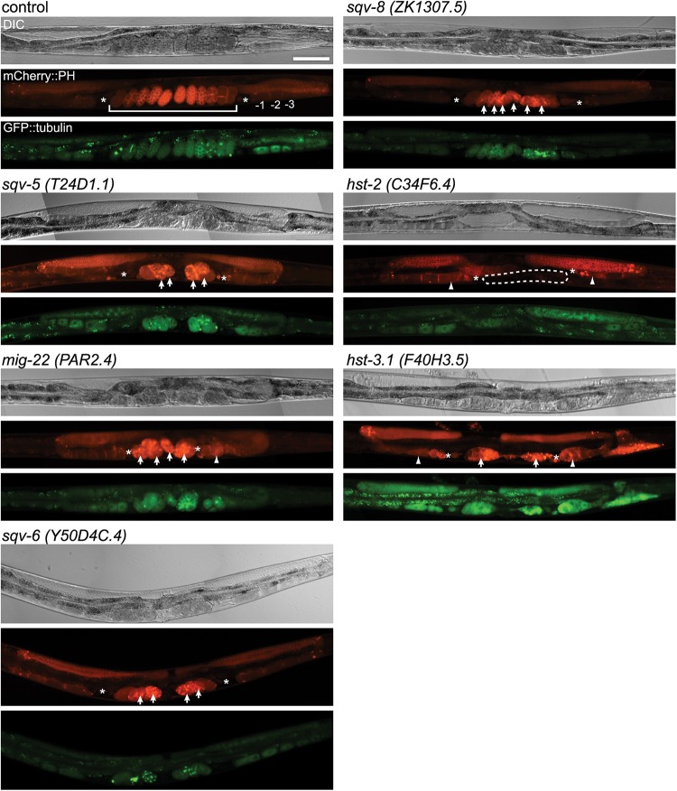
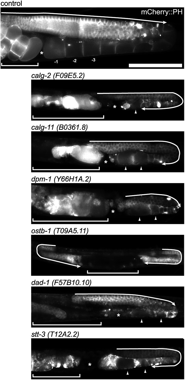
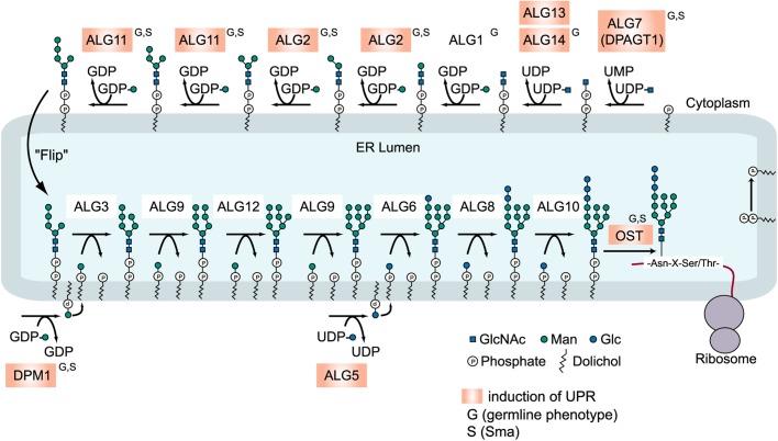
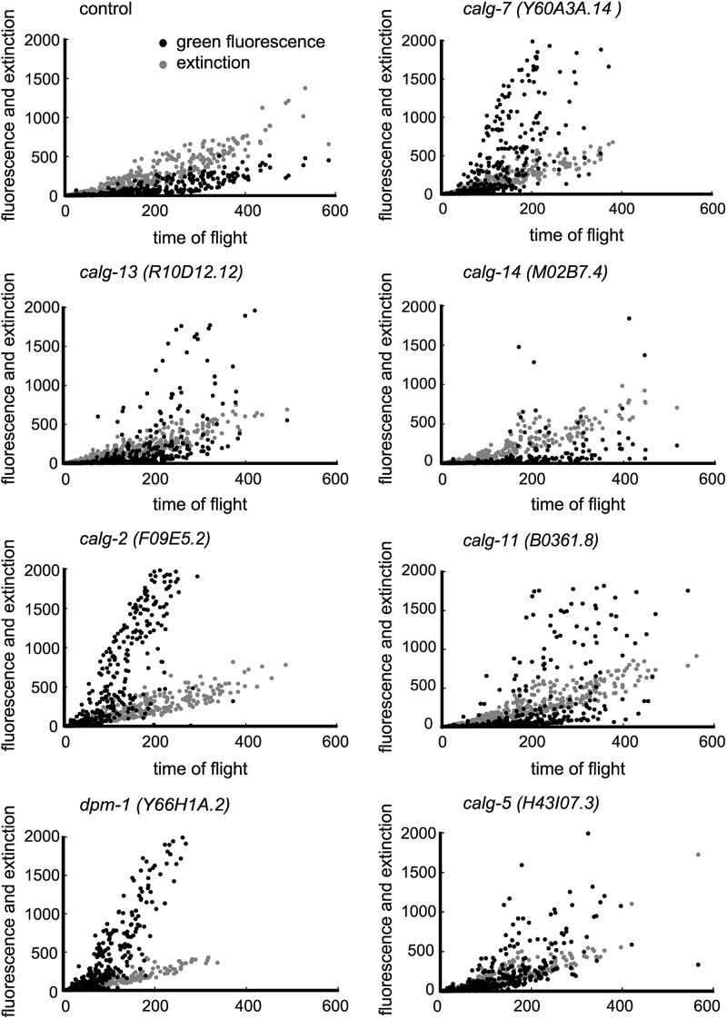
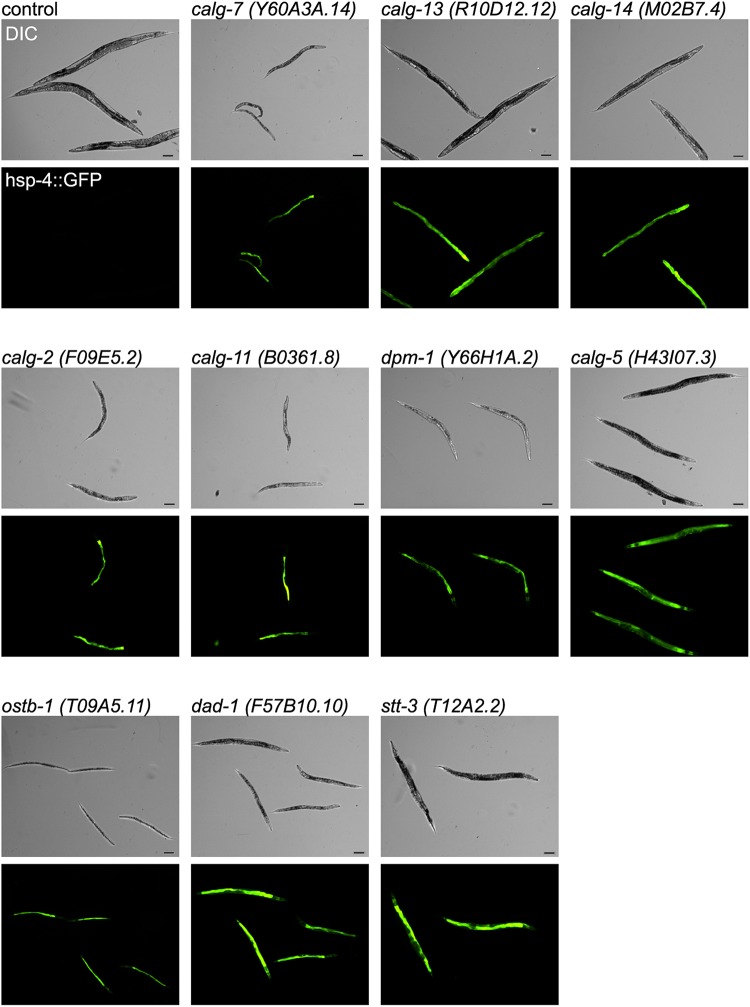
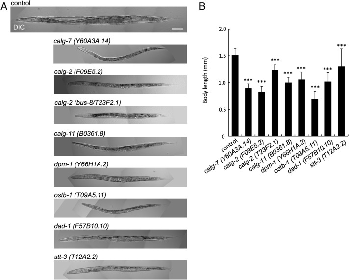
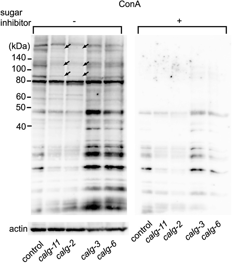
In this study, we selected 181 nematode glycogenes that are orthologous to human glycogenes and examined their RNAi phenotypes. The results are deposited in the Caenorhabditis elegans Glycogene Database (CGGDB) at AIST, Tsukuba, Japan. The most prominent RNAi phenotypes observed are disruptions of cell cycle progression in germline mitosis/meiosis and in early embryonic cell mitosis. Along with the previously reported roles of chondroitin proteoglycans, glycosphingolipids and GPI-anchored proteins in cell cycle progression, we show for the first time that the inhibition of the functions of N-glycan synthesis genes (cytoplasmic alg genes) resulted in abnormal germline formation, ER stress and small body size phenotypes. The results provide additional information on the roles of glycoconjugates in the cell cycle progression mechanisms of germline and embryonic cells.

Keywords: RNAi; alg genes; germline cell division; glycogene; oligosaccharyltransferase.


Figures

Fig. 1.
Fig. 1.
RNAi-mediated knockdown of several glycosyltransferases and sulfotransferases showed germline phenotypes. In each set of photographs, the upper panel shows the Nomarski DIC micrograph, the middle panel shows the PH domain detected by mCherry (red) and the bottom panel shows tubulin detected by gfp (green). In the mCherry::PH panel, the numbers indicate the normal oocytes, where −1 is the most proximal, and the brackets indicate the normal uterus (control); arrow: embryonic lethal (sqv-5, mig-22, sqv-6, sqv-8 and hst-3.1); arrowhead: oocyte morphology variant (mig-22, hst-2 and hst-3.1); dashed line: no eggs in uterus (hst-2), asterisks; spermatheca, scale bar: 100 μm.
Fig. 2.
Fig. 2.
RNAi-inactivation of glycosyltransferases involved in LLO synthesis showed germline phenotypes. The germline of the control shows the normal syncytium end (arrow). The knockdown of calg-2 and calg-11 resulted in the extension of the syncytial region over the loop of gonad (arrow). The oocyte number decreased, and the oocyte morphology became variant (arrowhead). dpm-1, dad-1 and stt-3 RNAi also produced abnormal oocytes (arrowheads), and ostb-1 and stt-3 RNAi resulted in expanded syncytium. The inhibition of ostb-1 resulted in no oocytes, a very short gonad and expanded syncytium (arrow) phenotypes. The RNAi of these glycosyltransferases involved in LLO (lipid-linked oligosaccharides) synthesis all showed a small gonad phenotype compared with the control. The PH domain was detected by mCherry. The brackets indicate the uterus region. The asterisk indicates the spermatheca. Scale bar: 100 μm.
Fig. 3.
Fig. 3.
Synthesis of dolicol-P-P-GlcNAc2Man9Glc3 in the ER of eukaryotes. A model for the eukaryotic biosynthetic pathway of lipid-linked oligosaccharides (LLOs) and its transfer to proteins. The glycogenes that showed an increase of hsp-4::GFP expression by RNAi are highlighted in orange, and the genes showing small body size (S) or germline phenotype (G) are indicated. ostb-1, dad-1 and stt-3 code subunits of OST.
Fig. 4.
Fig. 4.
RNAi of glycosyltransferases involved in LLO synthesis at the cytoplasmic side of ER resulted in the strong induction of hsp-4::GFP. The black dots represent hsp-4::GFP fluorescence, and the gray dots represent extinction, which provides a measurement of optical density of the organism. COPASTM Biosort was used for the analyses. X-axis: time of flight (length of each organism); Y-axis: intensity.
Fig. 5.
Fig. 5.
RNAi of C. elegans cytoplasmic alg gene orthologs involved in LLO synthesis and RNAi of OST subunit genes induced strong GFP fluorescence under fluorescence microscopy. In each of the micrographs, the upper panel shows the Nomarski DIC micrograph, and the bottom panel shows the fluorescence of the hsp-4::gfp (green). Scale bar: 100 μm.
Fig. 6.
Fig. 6.
RNAi-mediated knockdown of glycosyltransferases involved in LLO synthesis showed small (Sma) phenotype. (A) The RNAi-mediated inactivation of LLO synthesis genes resulted in small body size. Scale bar: 100 μm. (B) Body length of RNAi-treated animals. The error bar indicates the standard deviation. Control n = 28, calg-7 (Y60A3A.14) n = 33, calg-2 (F09E5.2) n = 20, calg-2 (bus-8/T23F2.1) n = 13, calg-11 (B0361.8) n = 21, dpm-1 (Y66H1A.2) n = 30, ostb-1 (T09A5.11) n = 25, dad-1 (F57B10.10) n = 36 and stt-3 (T12A2.2) n = 26. ***P ≤ 0.001.
Fig. 7.
Fig. 7.
Knockdown of calg-11 or calg-2 shows reduction of ConA staining. ConA staining was decreased in the RNAi-mediated inhibition of cytoplasmic alg genes (calg-2 and calg-11) compared with the control. The arrows indicate the bands showing strong reduction. This reduction was not detected under the inhibition of ER-lumen alg genes (calg-3 and calg-6). The blotted membrane of the same samples shown at the right panel was incubated with sugar inhibitor plus HRP-conjugated ConA (Plus sign), and the left membrane was incubated with HRP-conjugated ConA in the absence of the sugar inhibitor (minus sign), followed by ECL detection. The bottom panel shows the anti-actin staining of the same lanes of the same samples as a control.

References

    1. Ahringer J. Reverse Genetics. WormBook. 2006 doi:10.1895/wormbook.1.47.1. - DOI
    1. Akiyoshi S, Nomura KH, Nomura K. 2013. GlycoPOD RNAi protocols http://jcggdb.jp/GlycoPOD/protocolShow.action?nodeId=t205 , last accessed July 14, 2014.
    1. Bailey TL, Elkan C. Fitting a mixture model by expectation maximization to discover motifs in biopolymers. Proc Int Conf Intell Syst Mol Biol. 1994;2:28–36. - PubMed
    1. Berninsone PM. Carbohydrates and glycosylation. WormBook. 2006 The C. elegans Research Community, WormBook, doi/10.1895/wormbook.1.125.1, http://www.wormbook.org, last accessed July 14, 2014. - PMC - PubMed
    1. Bhattacharya R, Townley RA, Berry KL, Bulow HE. The PAPS transporter PST-1 is required for heparan sulfation and is essential for viability and neural development in C. elegans. J Cell Sci. 2009;122:4492–4504. - PMC - PubMed

Publication types

MeSH terms