. 2014 Aug 6:2:91.
doi: 10.1186/s40478-014-0091-0.

Fetal syringomyelia

Fetal syringomyelia

Anne Guo et al. Acta Neuropathol Commun. .

Abstract

We explored the prevalence of syringomyelia in a series of 113 cases of fetal dysraphism and hindbrain crowding, of gestational age ranging from 17.5 to 34 weeks with the vast majority less than 26 weeks gestational age. We found syringomyelia in 13 cases of Chiari II malformations, 5 cases of Omphalocele/Exostrophy/Imperforate anus/Spinal abnormality (OEIS), 2 cases of Meckel Gruber syndrome and in a single pair of pyopagus conjoined twins. Secondary injury was not uncommon, with vernicomyelia in Chiari malformations, infarct like histology, or old hemorrhage in 8 cases of syringomyelia. Vernicomyelia did not occur in the absence of syrinx formation. The syringes extended from the sites of dysraphism, in ascending or descending patterns. The syringes were usually in a major proportion anatomically distinct from a dilated or denuded central canal and tended to be dorsal and paramedian or median. We suggest that fetal syringomyelia in Chiari II malformation and other dysraphic states is often established prior to midgestation, has contributions from the primary malformation as well as from secondary in utero injury and is anatomically and pathophysiologically distinct from post natal syringomyelia secondary to hindbrain crowding.


Figures
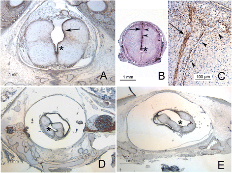
Figure 1
Figure 1
Syrinx morphology in Chiari II malformations. A Type 1 syrinx: the ventral portion of the cavity is ependymal lined (asterix), while the dorsal aspect is irregularly lined by a layer of hyperplastic glia (arrow). Despite the lack of dorsal ependymal lining, the impression that this is simply a wedge shaped extension of the more caudal dysraphism is difficult to escape. B Type 2 syrinx, extending in this section into the cervical cord, with a prominent paramedian and dorsal distribution. The arrow indicates the normal dorsal and median glial raphe and arrowheads the slit-like paramedian syrinx. This cavity was continuous with the dilated central canal (asterix) caudally. C Dorsal aspect of the same section as B at higher power, demonstrating glial processes lining a slit like cavity containing macrophages, with axonal tracts in the walls of the cavity. D and E: Same specimen, type 3 syrinx, demonstrating irregular extension of the syrinx both dorsolaterally (D) and ventrolaterally (E). The cavity marked by an asterix is partially lined by ependyma and is continuous with the central canal. All sections stained immunohistochemically for vimentin.
Figure 2
Figure 2
Spinal cord pathology associated with syringes in Chiari II malformations. A The anatomical boundary between syrinx and central canal may be indistinct, with syringes partially lined by ependyma (arrowheads) alternating with areas of glial hyperplasia (arrows, Immunohistochemistry for nestin). B Secondary changes in a spinal cord with syrinx: macrophages forming dense aggregates (arrow) and accumulating within the neuropil (arrowhead, immunohistochemistry for CD168). C H + E of same case as B, demonstrating aggregate of foamy macrophages. D Region of gliosis at area indicated by arrowhead in B (immunohistochemistry for alpha beta crystallin). E Vernicomyelia with foreign body macrophages (arrow). F Immunohistochemical reactivity of vernix to low molecular weight keratin (Cam 5.2).
Figure 3
Figure 3
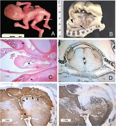
Spinal cord and syrinx pathology in OEIS. A OEIS in a midgestation fetus with large dorsal myelocystocele (arrowhead). B Longitudinal section of caudal spine and myelocystocele in a near term infant with OEIS. The dorsal sac (asterix) is lined by ependyma and continuous with the central canal of the spinal cord (arrow) which herniates dorsally. The dorsal elements of the neural canal are absent in the midline below the upper lumbar spine (arrowhead) and the conus is low lying. C Same specimen as (A), whole mounted axial section of spinal column at the point where the cord herniates through dorsally between the separated spinal laminae (arrowheads). The central canal (asterix) is dilated and irregular with a discontinuous lining. D Axial section of cervical spinal cord of same specimen as (A) demonstrating dorsal syrinx with lateral extensions separate from a mildly dilated central canal (immunohistochemistry for vimentin). E Transverse section through the cervical cord of the same fetus as (B), demonstrating an irregular dorsolateral cavity with adjacent increased staining for alpha beta crystallin (arrowheads), separate from normal sized central canal (asterix). F Same specimen and level as (E), stained for myelin basic protein, with near total loss of myelin staining adjacent to the syrinx cavity.
Figure 4
Figure 4
Syringes in Meckel Gruber and pyopagus twins. A Cross section of cervical spinal cord, Meckel Gruber with an occipital encephalocele. A small dorsal median cavity (arrow) is continuous ventrally with the dorsal glial raphe, but does not appear to connect to the mildly dilated and dorsally elongated central canal in this plane. B Same case, thoracolumbar cord: large dorsal syrinx with irregular lateral extensions (A and B: Immunohistochemistry for Vimentin). C Wall of syrinx in (B), with scattered histiocytes reacting positively for Iron (arrows, Perl’s iron stain). D Sacral spinal cords of pyopagus twins stained for alpha beta crystallin: one cord is a hemicord with a collapsed and dilated central canal (asterix). Both cords share a thin fibrolipomatous sheath within a single dural sac. More rostrally, the lumbar spinal cords of each twin separated and had distinct dural sacs within a common neural canal. E Cross section of thoracolumbar spinal of the cord oriented as the superior cord in D demonstrating a dorsally elongated central canal lined by ependyma and extending between the posterior columns. F The abnormality in (E) is duplicated in the thoracolumbar cord of the other twin.

References

    1. Driver CJ, Volk HA, Rusbridge C, Van Ham LM. An update on the pathogenesis of syringomyelia secondary to Chiari-like malformations in dogs. Vet J. 2013;198(3):551–9. doi: 10.1016/j.tvjl.2013.07.014. - DOI - PubMed
    1. Klekamp J, Samii M. Syringomyelia: Diagnosis and Treatment. Heidelberg: Springer-Verlag Berlin; 2002.
    1. Levine DN. The pathogenesis of syringomyelia associated with lesions at the foramen magnum: a critical review of existing theories and proposal of a new hypothesis. J Neurol Sci. 2004;220(1–2):3–21. doi: 10.1016/j.jns.2004.01.014. - DOI - PubMed
    1. Milhorat TH, Capocelli AL, Anzil AP, Kotzen RM, Milhorat RH. Pathological basis of spinal cord cavitation in syringomyelia: analysis of 105 autopsy cases. J Neurosurg. 1995;82:802–812. doi: 10.3171/jns.1995.82.5.0802. - DOI - PubMed
    1. Milhorat TH1, Miller JI, Johnson WD, Adler DE, Heger IM. Anatomical basis of syringomyelia occurring with hindbrain lesions. Neurosurgery. 1993;32(5):748–54. doi: 10.1227/00006123-199305000-00008. - DOI - PubMed

MeSH terms

Supplementary concepts